Gram Staining: Definition, Procedure, Facts, Topics, Diagram
What Is Gram Staining?
In microbiology, Gram stain or Gram’s method is a method employed for distinguishing bacteria based on the characteristics of the cell wall. Invented in 1884 by Danish scientist Dr Hans Christian Gram, it entails the use of crystal violet dye, iodine, alcohol and safranin. Gram staining is used principally to distinguish bacteria into two groups namely the Gram positive and the Gram negative. Septum thickness is thicker in Gram-positive bacteria compared to Gram-negative bacteria. Gram-positive bacteria retain the violet stain because of the thick layer of peptidoglycan while Gram-negative bacteria lose the stain due to the thin layer of peptidoglycan and a membranous layer surrounding it.
Latest: NEET 2024 Paper Analysis and Answer Key
Don't Miss: Most scoring concepts for NEET | NEET papers with solutions
New: NEET Syllabus 2025 for Physics, Chemistry, Biology
NEET Important PYQ & Solutions: Physics | Chemistry | Biology | NEET PYQ's (2015-24)

This differentiation is crucial in microbiology, in the assessment of pathogens, and assessment of bacterial characteristics or treatment regimes. Hans Christian Gram’s contribution to microbiology was to provide a profound new concept by developing an amazingly effective yet much-needed method that remains the key for bacterial categorisation in clinical diagnostic laboratories and research centres across the globe.
Principles Of Gram Staining
The principles of gram staining are listed below
Cell Wall Composition
Explanation of Gram-positive and Gram-negative bacteria.
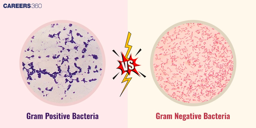
Gram-positive bacteria have many layers of peptidoglycan envelope in their cell wall, crystal violet dye which is used in gram staining attaches to them therefore they appear purple. By comparison, carbohydrates in Gram-negative bacteria are thinner and surrounded by an outer membrane which is composed of lipopolysaccharides, and after staining with safranin, it turns pink.
Structural differences in peptidoglycan layers
The purification of gram-positive bacteria has a resistant and homogeneous layer of peptidoglycan that retains the crystal violetiodine complex explaining why they take the purple stain. However, due to the thin layer of peptidoglycan in the middle of the outer and inner membranes present in the gram-negative bacteria the crystal violet stain is decolorised during the decolorisation step and the safranin counter stain is then taken up hence appearing pink or red.
Staining Procedure
Stepbystep process:
Fixation: These include; In the first step, heat fixation is done whereby the bacterial smear is heated causing the bacteria to be firmly attached to the slide.
Crystal violet staining: The slide is then flooded with crystal violet a primary stain that will colour all the cells purple to enhance visibility.
Iodine treatment: The slide is treated with iodine solution for it to form a complex with crystal violet within the cells to increase its stain retention.
Alcohol decolourisation: An alcohol or ethanol wash is applied to wash out the stain from cells that fail to retain the stain and hence become colourless.
Counterstaining with safranin: Last, of all, the slide is stained with safranin that dyes decolourized cells a pink colour as a contrast to the green colour of PI. This step enables the differentiation of various bacteria by measuring the amount of stain that each would retain.
Interpretation of Results
Grampositive bacteria
These bacteria look purple or blue under the lens since they retain the crystal violet as the peptidoglycan layer in the cell wall is thicker.
Gramnegative bacteria
These bacteria are pink or red in specimens that are stained with gram because, during the decolourisation step in gram staining, these bacteria lose the crystal violet stain but retain the safranin counterstain and due to the thin layer of peptidoglycan in their cell wall and the additional outer membrane.
Diagram of Gram-positive and Gram-negative bacteria post staining.
Importance Of Gram Staining
The importance of Gram Staining is discussed below:
Clinical Applications In Diagnosis Of Bacterial Infections
In clinics, Gram staining plays a significant role in the proper identification of Gram-positive and negative bacteria and the choice of antibiotic to the characteristics of bacteria found in a sample.
Role In Research, Epidemiology, And Antibiotic Susceptibility Testing
In research, this staining technique assists the classifiers in the identification of bacteria in particular their morphology and how they are pathogenic. It is also used in epidemiology for example in revealing the behaviour of bacterial infections in a given population and in antibiotic susceptibility testing where one is bound to determine the efficiency of the antibiotics against different types of bacteria.
Recommended video for "Gram Staining"
Frequently Asked Questions (FAQs)
Gram staining locates bacteria through differential characteristics of cell wall and they are either Gram-positive or Gram-negative.
Another reason why this stain is crucial in microbiology is because it can be useful in the identification of bacteria, treatment strategies on patients and giving aspects of the bacterial characteristics like form and response to antibiotics.
However, it has to be noted that Gram staining cannot differentiate all types of bacteria since some bacterial species may not conform for classification to Gram staining due to the differences in their cell wall structure.
Since the procedure of Gram staining is complex if the right procedure is not followed the bacteria might not be stained or misclassified. This could, of course, impact diagnostic and therapeutic conclusions in some cases in the clinic or hospital.
Yes, Gram staining is also used in the identification and characterisation of bacteria in environmental microbiology and food microbiology found in samples of food products.
Also Read
28 Sep'24 09:36 PM
19 Sep'24 12:35 PM
19 Sep'24 12:30 PM
19 Sep'24 12:29 PM
19 Sep'24 12:13 PM
19 Sep'24 12:01 PM
19 Sep'24 11:39 AM
Articles
Download Careers360 App's
Regular exam updates, QnA, Predictors, College Applications & E-books now on your Mobile

300M+
Students

36,000+
Colleges

550+
Exams

1500+
E-Books

16000+
Certifications
