Cardiac Cycle: Definition, Diagram, Steps and Facts
The cardiac cycle is the sequence of events that occur during one heartbeat, ensuring blood is pumped efficiently throughout the body. It consists of two main phases: systole (contraction) and diastole (relaxation) of the heart chambers. This cycle maintains the rhythmic flow of oxygenated and deoxygenated blood, playing a crucial role in the circulatory system. In this article, the Cardiac Cycle, Phases of the Cardiac Cycle, Heart Sounds, Duration of the Cardiac Cycle, and Conditions and Diseases are discussed. Cardiac Cycle is a topic of the chapter Body Fluids and Circulation in Biology.
NEET 2025: Mock Test Series | Syllabus | High Scoring Topics | PYQs
NEET Important PYQ's Subject wise: Physics | Chemistry | Biology
New: Meet Careers360 B.Tech/NEET Experts in your City | Book your Seat now
- What is the Cardiac Cycle?
- Phases of the Cardiac Cycle
- Heart Sounds
- Duration of Cardiac Cycle
- Conditions and Diseases

What is the Cardiac Cycle?
The cardiac cycle is the process of a single series of heartbeats, which describe the contraction and relaxation of the heart’s chambers to pump blood to the body tissues. This cycle is very important for the continuous circulation of blood where oxygen and nutrients are taken to the tissues and wastes are removed. The cardiac cycle includes several key phases: systole which essentially involves the contraction of the heart to pump blood to the arterial system and diastole phase which is the period of relaxation of the heart after it has filled the self with blood. The knowledge of the cardiac cycle is important in any practice when it comes to the understanding of the function of the heart in the circulatory system which is a vital part of diagnosing cardiovascular disorders.

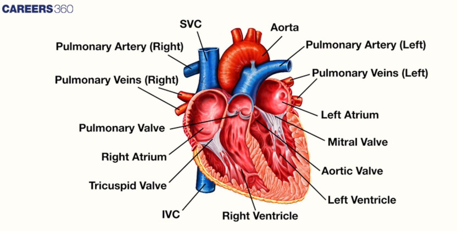
Detailed labelled diagram of the Heart
Phases of the Cardiac Cycle
The phases of the cardiac cycle are listed below-
Atrial Systole
Systole atrial is a phase where the atrial muscles contract.
This contraction forces blood into the ventricles, which finalises the submission of that chamber and raises the pressure in the ventricle.
The contraction of the atria makes sure that the ventricles are filled with blood in readiness for the next phase of pumping.
Ventricular Systole
This phase commences with the myocardium of ventricles contracting without an alteration of the volume due to the shutting of the valves.
Thus, the pressure inside the ventricles starts to rise rapidly, becoming higher than the pressure inside the atria.
When the pressure in the ventricles becomes higher than in the aorta and the pulmonary arteries this opens the semilunar valves and then the blood is expelled from the ventricles into the arteries.
Diastole
After ventricular ejection, both ventricles will be in the process of relaxation while the ventricular volumes remain the same and the semilunar valves close while atrioventricular valves are still closed.
After the valves of the atria are closed, the ventricles start to decompress, and the pressure within them becomes lower than the pressure in the atria, therefore, the atrioventricular valves open and, hence, the blood from the atria flows into the ventricles and the cycle is complete.
Also Read-
Heart Sounds
The heart sounds are discussed below-
S1(First Heart Sound)
It is due to the closure of atrioventricular (AV) valves; tricuspid and mitral resulting from the start of ventricular systole. This sound known as the “lub” is the first phase of the contraction of the heart and is important for the first sound of the heartbeat.
S2(Second Heart Sound)
These are created by the shutting of the semilunar valves, pulmonary and aortic, at the end of the ventricular systole. This sound known as ‘dub’ marks the point when the contraction phase of the heart stops and the diastole, which is the relaxation of the ventricles starts.
Additional Heart Sounds
The additional heart sounds are:
S3 (Third Heart Sound)
Mainly said after S2, is caused by the fast filling of ventricles during the early phase of diastole. This is very common, especially in the youthful and healthy populations, however, the older population may experience heart failure or volume overload. It is referred to as “ventricular gallop. ”
S4(Fourth Heart Sound)
Found immediately before S1 and is related to atrial systole in an attempt to force blood into a stiff or hypertrophied ventricle. This sound is known as the ‘atrial gallop’ and may occur in ailments such as hypertension as well as aortic stenosis.
Murmurs
Heart murmurs are just sounds that are produced by the turbulent blood flow in the valves or in the heart chambers.
Valvular diseases can stem from valvular lesions, where the valves are stenotic, regurgitant or there is an abnormal connection between the atria and ventricles.
Systolic Murmurs: Happen between S1 and S2 and are linked to pathologies that comprise aortic stenosis and/or mitral regurgitation.
Diastolic Murmurs: Occur between S2 and the following S1, and may be due to mitral stenosis or aortic regurgitation.
Continuous Murmurs: Lasts throughout the cardiac cycle, that is those seen in conditions like patent ductus arteriosus.
Duration of Cardiac Cycle
A normal person's heart beats at a rate of 72 beats per minute. Thus, one cardiac cycle's duration can be computed as follows:
1/72 beats per minute equals.0139 beats per minute.
Each cardiac cycle will last 0.8 seconds at a heart rate of 72 beats per minute.
The following lists the duration of the various cardiac cycle stages:
The duration of atrial systole is around 0.1 seconds.
The duration of ventricular systole is around 0.3 seconds.
The duration of atrial diastole is around 0.7 seconds.
Ventricular diastole lasts for almost half a second.
Conditions and Diseases
The diseases related to defective cardiac cycle are:
Hypertension
Hypertensive disease and hypertension in particular worsen the demand on the heart and radical changes in the cardiac cycle by increasing afterload and thus negatively affect the heart muscle over time.
Heart Failure
A state, in which stroke volume and heart rate are endangered due to the failure of the heart to pump blood efficiently. This can alter the rhythms of the normal cardiac cycle and that can cause such symptoms like fatigue and fluid retention.
Valve Disorders
Diseases such as stenosis or regurgitation relate to the valves and influence the circulating movement of blood through the heart and its capacity to fulfil the work of the cardiac cycle. Heart sounds become abnormal and cardiac output is decreased as a result of poor circulation.
Also Read-
| Facts about Blood | Facts about Blood Group |
| Types of Blood Cells | Blood coagulation |
| ABO Blood Group | Circulatory System |
Recommended video for Cardiac Cycle
Frequently Asked Questions (FAQs)
Atrial Systole: Chambers agree to take Atria contract to fill ventricles.
Ventricular Systole: Comprises are Isovolumetric Contraction and Ventricular Ejection.
Diastole: This consists of Isvolumetric relaxation and ventricular filling Differences between the right and left sides of the heart.
The sequence is the atrial systole, which fills the ventricles, ventricular systole, which ejects blood, and the diastolic periods for refilling the ventricles.
It describes the circulation of Blood through the Heart and the rest of the body to supply oxygen and nutrients and remove waste products.
It is controlled by the heart conduction system: SAN and AVN and modulated by ANS and hormones.
Atrial Systole: Atria contract to push blood into ventricles.
Ventricular Systole: Cavity sends blood out into arteries after the ventricles close.
Also Read
29 Nov'24 01:19 PM
27 Nov'24 07:39 PM
27 Nov'24 07:15 PM
27 Nov'24 05:11 PM
26 Nov'24 08:14 PM
26 Nov'24 06:50 PM
26 Nov'24 05:51 PM
26 Nov'24 04:44 PM
26 Nov'24 03:52 PM
26 Nov'24 02:55 PM
Articles

Student Community: Where Questions Find Answers