Chromosomes: Definition, Types, Structure and Function
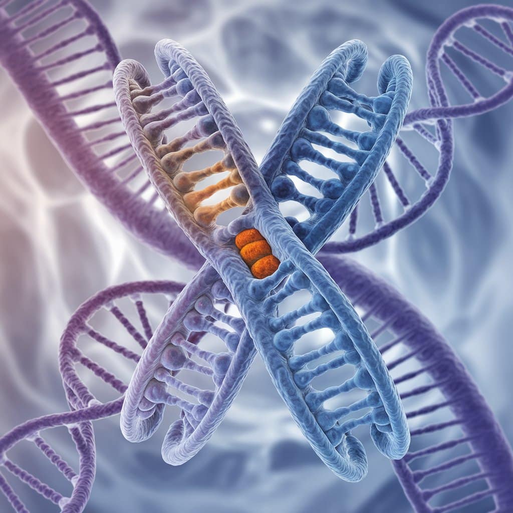
Chromosomes Definition: Chromosomes are thread-like structures in cells that carry genetic information. They ensure genetic material is passed from parents to offspring and control traits. This topic is from the class 12 chapter Molecular Basis of Inheritance in Biology. This article includes information about the structure of chromosomes, types of chromosomes, how many chromosomes are present in endosperm, what chromosomes are and the function of chromosomes
NEET 2025: Mock Test Series | Syllabus | High Scoring Topics | PYQs
NEET Important PYQ's Subject wise: Physics | Chemistry | Biology
New: Meet Careers360 B.Tech/NEET Experts in your City | Book your Seat now
- What are Chromosomes?
- Discovery of Chromosomes
- Role of Chromosomes in Genetics
- Composition of Chromosomes
- Types of Chromosomes
- How Many Chromosomes in Humans?
- Chromosomal Disorders
- Cell Division and Chromosomes
- What is the Function of Chromosomes?
- Chromosomes in Evolution and Speciation

What are Chromosomes?
Chromosomes are long thread-like structures found in the eukaryotic cell nucleus composed of DNA and Proteins. This chromosome plays a critical role in the storage and transmission of genetic information generation after generation. It is important in inheritance because it ensures accurate copying and distribution of DNA at the time of cell division.
Discovery of Chromosomes
Matthias Schleiden and Theodor Schwann laid the foundation of cell theory, establishing that all living organisms are composed of cells, which are the basic units of life.
Gregor Mendel conducted pioneering work on heredity, laying the groundwork for understanding genetic inheritance through his experiments with pea plants.
Thomas Hunt Morgan studied fruit fly chromosomes, providing crucial evidence for the chromosomal theory of inheritance and demonstrating that genes are located on chromosomes.
Walter Sutton and Theodor Boveri developed the chromosomal theory of inheritance, explaining how chromosomes are responsible for gene regulation and the inheritance of traits.
Also Read:
- MCQ Practice on Chromosomal Mutations
- Molecular Basis of Inheritance
- Mendelian Disorders
- Chromosomal Abnormalities
Role of Chromosomes in Genetics
Chromosomes primarily work in genetics as they contain the genetic information in genes that give an organism its characteristics and control gene expression.
It plays a crucial role in proper inheritance, gene expression, and the evolutionary process to ensure that the information to be passed from one generation to another is materialised through the proper transmission of genetic information.
Composition of Chromosomes
Chromatin is a DNA-protein complex in the interphase nucleus of the cell, presenting an amorphous, decondensed appearance.
At the time of cell division, however, chromatin condenses into visible, differentiated chromosomes so that the DNA can be separated in a highly ordered and precise manner.
Components of Chromosomes
DNA: The DNA encodes the genetic blueprint of the organism.
Histone Proteins: Histone proteins package and order the DNA in the form of organized units called nucleosomes; they help to organize and control the genetic material.
Non-Histone Proteins: They play a variety of functions including chromosome maintenance and gene regulation.
Centromere: It is the middle part of a chromosome where sister chromatids are held and also plays a critical role in the movement of chromosomes during cell division
Chromatids: Duplicated halves of the same chromosome divided during mitosis and meiosis
Telomeres: Protective caps that feature at the ends of the chromosomes that help prevent their degradation and fusion with other chromosomes.
Kinetochore: A complex of proteins attached to the centromere that attaches spindle fibres to the chromosomes during cell division.
Types of Chromosomes
Based on the Number of Centromeres:
Monocentric: It has only one centromere.
Dicentric: Contain two centromeres.
Acentric: Lack of centromere.
Holocentric: Present with centromeres at all other places on the entire chromosome length.
Based on the Position of the Centromere:
Metacentric: The centromere lies in the centre. Results in both arms of the same length.
Submetacentric: The centromere is found off-centre. Results in unequal length of the two arms.
Acrocentric: Centromere near one end, so a very short p arm and long q arm.
Telocentric: Centromere at the end of the chromosome, thus no p arm.
How Many Chromosomes in Humans?
Introduction to the number of chromosomes in humans:
Humans have 46 chromosomes in total, which occur in 23 pairs, 22 pairs are autosomes, and the remaining pair consists of sex chromosomes.
Autosomes vs. Sex Chromosomes
Autosomes: These include the chromosome pairs 1-22. It has similar characteristics in both sexes and determines most of the traits of an individual.
Sex Chromosomes: This includes an X and Y chromosome, responsible for determining the sex of an individual. Females possess the XX chromosomes, while males carry the XY chromosomes.
Chromosomes vs Chromatids
Chromosomes are single, thread-like structures that contain DNA. Chromatids are identical halves of a duplicated chromosome, connected at the centromere, and separated during cell division.
Chromosomal Disorders
Numerical Abnormalities (Aneuploidy) are:
Trisomy 21: Also known as Down syndrome. This is the presence of an extra copy of chromosome 21.
Monosomy X: That is Turner syndrome, a condition of having only one X chromosome in females.
Klinefelter Syndrome: The condition in males where there is an extra X chromosome (XXY) that affects sexual development and fertility.
Structural Abnormalities:
Deletions: A chromosome segment missing from the genetic material of a person, which may cause genetic disorders.
Duplications: Doubling of a chromosome segment that might be the reason for many genetic imbalances.
Inversions: The reversal of a segment of a chromosome, which can impact gene function.
Translocations: Exchange of a part of a chromosome between two non-homologous chromosomes. Most often, such an exchange causes genetic disorders or cancer.
Cell Division and Chromosomes
The role of chromosomes in cell division is explained as:
Mitosis
It ensures that daughter cells both contain the same number of chromosomes.
Prophase: Chromosomes condense and create the mitotic spindle.
Metaphase: Chromosomes align along the equatorial plane of the cell.
Anaphase: Sister chromatids separate to opposite poles.
Telophase: Chromosomes start to de-condense, and nuclear membranes reform.
Cytokinesis: The cell membrane pinches in to divide the cell into two.
Meiosis
Reduction Division for Gametes: Meiosis reduces the chromosome number by half to produce gametes (sperm and eggs).
Meiosis I: Homologous chromosomes are separated into different cells, reducing the chromosome number by half.
Meiosis II: Sister chromatids are separated into different cells, resulting in four genetically diverse gametes.
Crossing Over: It occurs in Prophase I, that is, the genetic materials are exchanged between homologous chromosomes and thereby increase the genetic variation.
What is the Function of Chromosomes?
The function of chromosomes are given below:
Storage of Genetic Information
The chromosomes take, carry, and transfer genetic information from the DNA. This ensures that the genetic instructions regarding growth, development, and function would be conserved and passed on to the next generation.
Gene Regulation
Chromosomes regulate gene expression by controlling the accessibility of DNA to the transcription machinery.
Cell Cycle Regulation
Chromosomes are involved in various cell cycle phases, DNA replication during the S phase and chromosome segregation during both mitosis and meiosis.
Methods for Chromosome Analysis
The methods of chromosome analysis are:
Karyotyping
Karyotyping is the process of aligning chromosomes in a standardized manner to detect abnormalities of chromosomes and diagnose genetic diseases.
Fluorescence In Situ Hybridisation (FISH)
FISH utilises fluorescent probes to hybridise with specific DNA sequences on chromosomes to facilitate the visualisation of chromosomal anomalies and gene localisation.
Chromosome Banding
G-banding: It results in the generation of banding due to the use of Giemsa stain in staining, which further aids in the identification of the chromosomes
Q-banding: It utilises quinacrine mustard dye to highlight the different chromosome regions
R-banding: One can visualise reverse G-banding patterns by using heat and staining
Chromosomes in Evolution and Speciation
Chromosomal alterations create genetic diversity and evolve the species into various forms, thus affecting reproductive isolation.
Chromosomal Mutations and Speciation
Examples: new species can arise due to duplication, deletion, and translocation of chromosomes that change the gene content and function. These changes may, at times, result in reproductive barriers.
Importance of Chromosomes in Medical Research
The importance of chromosomes in medical research is explained below:
Genetic Engineering and Gene Therapy
Chromosome manipulation is very important in the development of treatments for genetic diseases, including gene therapy to correct defective genes.
Cancer Research
The mechanism of cancer can be understood through chromosomal abnormalities. Targeted therapy was developed as a result of understanding the Philadelphia chromosome associated with chronic myeloid leukaemia.
Also Read:
Frequently Asked Questions (FAQs)
Chromosomes are considered to consist of DNA tightly coiled around proteins known as histones that give a structure called chromatin.
Humans have 46 chromosomes in each somatic cell, in 23 pairs.
Chromosomal disorders result from abnormalities in chromosomal number or structure, including Down syndrome, Turner syndrome, and Klinefelter syndrome.
Chromosomes carry genetic information that is crucial for controlling inheritance, activating genes, and separating cells.
Chromosomes are then inherited from each parent, half through the egg from the mother and half through the sperm from the father.
The endosperm in flowering plants contains three sets of chromosomes (3n) due to the process of double fertilization.
Also Read
29 Nov'24 09:31 AM
19 Nov'24 09:26 AM
18 Nov'24 06:45 PM
18 Nov'24 09:29 AM
18 Nov'24 09:18 AM
18 Nov'24 09:01 AM
18 Nov'24 08:37 AM
16 Nov'24 03:45 PM

