

Diagram of Eye: Pictures, Images, Stock Photos
A labelled diagram of the human eye helps learners easily understand the anatomy of one of the most complex sensory organs. Its major external and internal structures—cornea, lens, iris–pupil system, retina, and optic nerve—work together to capture and process visual information. This guide explains all parts of the eye with diagram cues, functions, and NEET-ready MCQs.
This Story also Contains
- Introduction — Anatomy via Eye Diagram
- External Structures of the Human Eye
- Internal Structures of the Human Eye
- Diagram of Eye NEET MCQs (With Answers & Explanations)
- Recommended Video on Diagram of Eye
Introduction — Anatomy via Eye Diagram
The human eye is a complex organ specialized for vision, capturing incoming light and converting it into electrical signals interpreted by the brain. A labelled diagram helps visualize the spatial arrangement of structures such as the cornea, lens, retina, and optic nerve. It also makes it easier to understand how these parts work together in facilitating vision.
External Structures of the Human Eye
The extrinsic structures act to protect the eye and facilitate its function.
Eyelids
The eyelids protect the eye from debris and control the amount of light falling on the eye.
Conjunctiva
It is a thin membrane covering the front of the eye and lining the inner eyelids.
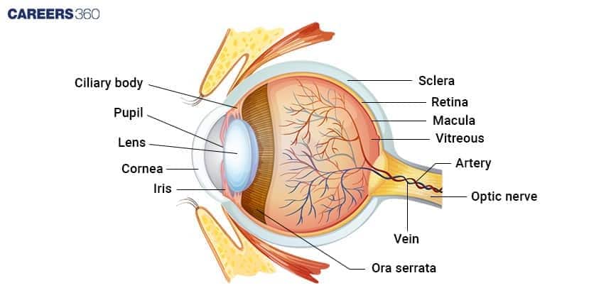
Internal Structures of the Human Eye
The structures inside the eye complete the vision.
Cornea
The transparent, curved surface at the front of the eye refracts light.
Iris And Pupil
The iris controls the diameter of the pupil, which regulates the amount of light reaching the eye.
Lens
Flexible, transparent, focusing light onto the retina.
Retina
The inner one with photoreceptor cells which detect the light—rods and cones, which transmit it to the brain.
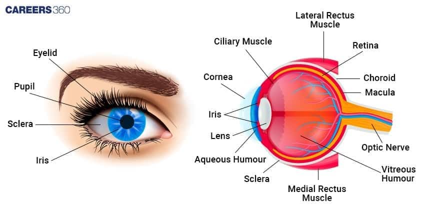
Diagram of Eye NEET MCQs (With Answers & Explanations)
Important questions asked in NEET from this topic are:
External and internal anatomy of human eye
Function of human eye
Practice Questions for NEET
Q1. The transparent lens in the human eye is held in its place by
Smooth muscles attached to the iris
Ligaments attached to the iris
Ligaments attached to the ciliary body
Smooth muscles attached to the ciliary body
Correct answer: 3) Ligaments attached to the ciliary body
Explanation:
The middle layer of the eye, also known as the vascular coat, consists of several key structures. In front of the lens, the pupil is the aperture surrounded by the iris, and its diameter is regulated by the muscle fibers of the iris. The transparent crystalline lens of the eyeball is held in place by ligaments attached to the ciliary body, which also forms a pigmented, opaque structure known as the iris, the visible colored portion of the eye. The choroid layer, which forms the posterior two-thirds of the eyeball, is thin but becomes thicker in the anterior region to form the ciliary body. The choroid contains numerous blood vessels and appears bluish, providing nourishment to the eye. Therefore, the transparent lens in the human eye is held in place by ligaments attached to the ciliary body.
Hence, the correct answer is option 3) ligaments attached to the ciliary body.
Q2. Which of the following statements is correct?
The cornea is an external, transparent and protective proteinaceous covering of the eye - ball
Cornea consists of dense connective tissue of elastin and can repair itself
Cornea is a convex, transparent layer which is highly vascularised.
Cornea consists of a dense matrix of collagen and is the most sensitive portion of the eye.
Correct answer: 1) The cornea is an external, transparent and protective proteinaceous covering of the eye - ball
Explanation:
The cornea is the outermost, transparent layer at the front of the eye that protects the eye's interior. It is made primarily of proteins, particularly collagen, which gives it a tough and protective structure. The cornea allows light to enter the eye and plays a key role in focusing vision by refracting light. Its transparency and curvature are essential for proper vision.
Hence the correct answer is option 1) The cornea is an external, transparent and protective proteinaceous covering of the eye - ball.
Q3. The choroid layer is thin over the posterior two-thirds of the eyeball, but it becomes thick in the anterior part to form the
Iris
Cornea
Pupil
Ciliary body
Correct answer: 4) Ciliary body
Explanation:
The front portion of the eyeball, which forms the ciliary body, has a thicker choroid layer than the posterior two-thirds. The ciliary muscles, which regulate the lens's shape for focusing, are found in the ciliary body, which also produces aqueous humor. Additionally, it is connected to the iris, which controls how much light enters the eye.
Hence, the correct answer is option 4)Ciliary body.
Also Read:
Recommended Video on Diagram of Eye
Frequently Asked Questions (FAQs)
The eyelids protect the eye from dust and other particles, thus regulating the entry of light into it.
The pupil regulates the amount of light that might get into the eye.
It bends light which passes through into the eye.
The shape of the lens changes to focus light into the retina.
The light is picked by the retina and then transmitted into the brain to process the vision.
