Diagram Of Neuron: Detailed Explanations, Structure & Function
A neuron diagram provides a clear visual representation of the structure and parts of a nerve cell responsible for receiving, processing, and transmitting information. Understanding each labelled part—dendrites, axon, soma, myelin, nodes, synaptic terminals—is essential for Class 11 Biology and NEET. This guide covers neuron structure, types, functions, diagrams, FAQs, and NEET-level MCQs.
This Story also Contains
- What Is a Neuron?
- Labelled Neuron Diagram
- Structure of a Neuron
- Types of Neurons
- How Neurons Work – Signal Transmission
- Neuron-Related Disorders
- Neurons NEET MCQs (With Answers & Explanations)
- Recommended Video on ‘Diagram of Neuron’

What Is a Neuron?
Neurons are the basic building blocks of the brain and nervous system and serve to receive sensory input, send motor commands to the muscles, and modify and transmit electrical signals throughout the body. Each neuron is specialised to perform transmission of information to other nerve cells, muscle, or gland cells. They conduct all the activities taking place in the nervous system: reflex actions, perceiving, thinking, memorising, and the performance of voluntary movements as well as involuntary movements.
Neurons communicate with each other through synapses by changing electrical impulses into a chemical form to ensure efficient and correct transmission of the signal. The structure and functions of neurons are very important in understanding the activities going on in the brain and in treating neurological disorders.
Labelled Neuron Diagram
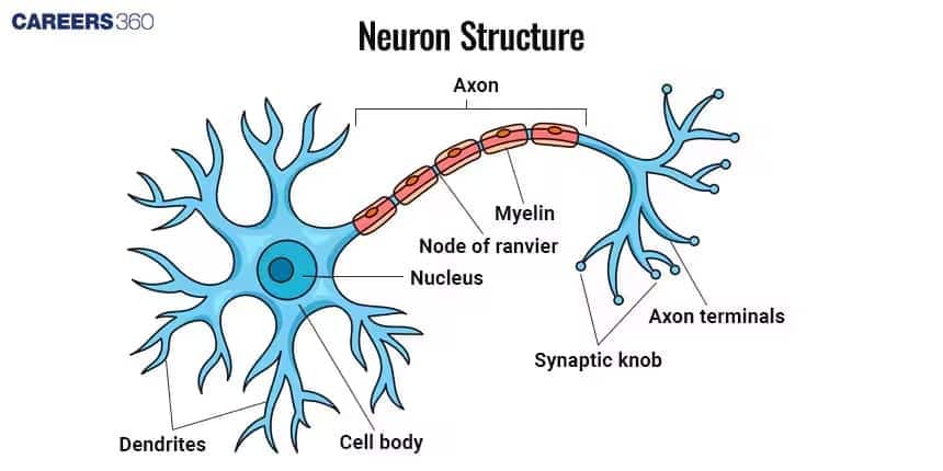
Given below is the simple neuron diagram with its parts:

Structure of a Neuron
The neuron structure contains several primary parts that make the cell functional. These parts include:
Cell Body (Soma)
Contains a nucleus and organelles.
Processes information and maintains cell health.
Dendrites
Branch-like structures which receive signals from other neurons.
Increase surface area for signal reception.
Axon
Long, slender projection which transmits signals away from the cell body.
Axons come in different lengths depending on the type of neuron.
Axon Terminals
These are the endings of the axon that connect to other neurons or muscle cells.
Release neurotransmitters to communicate with other cells.
Myelin Sheath
It is the insulating membrane around axons.
It increases the speed of nerve impulse transmission.
Nodes of Ranvier
Gaps in the myelin sheath
Saltatory conduction quickly transmits the signal.
Types of Neurons
Neurons are classified based on the structure and functionality of the neuron. They are classified into:
Sensory Neurons
It carries sensory information from receptors to the central nervous system.
Example: Neuron in the skin which detects touch.
Motor Neurons
Carry commands from the central nervous system to muscles.
Example: Neuron which causes muscle contraction
Interneurons
Connect sensory and motor neurons within the central nervous system.
Example: Neurons in the spinal cord that relay signals between sensory and motor neurons.
How Neurons Work – Signal Transmission
Neurons transmit signals through action potentials and chemical communication at synapses.
Action Potential
An action potential is an electrical impulse generated by changes in membrane ion permeability.
Synapse Structure
The synapse consists of the presynaptic terminal, which releases neurotransmitters.
The synaptic cleft, which acts as the gap between neurons.
The postsynaptic membrane, which receives and responds to the chemical signal.
Neurotransmitters
Dopamine: Helps regulate reward, movement, and mood.
Serotonin: Influences emotions, sleep cycles, and overall well-being.
Acetylcholine: Enables muscle contraction and is essential for learning and memory.
Neuron-Related Disorders
Several neurological diseases are claimed to be connected to the failure of neurons.
Alzheimer’s Disease
A condition characterised by the death of neurons, and the consequences are memory loss and cognitive declines
Parkinson’s Disease
Involves the loss of dopamine-producing neurons, affecting movement and coordination.
Multiple Sclerosis
An autoimmune disorder that attacks the myelin sheath, disrupting neural communication.
Neurons NEET MCQs (With Answers & Explanations)
Important questions asked in NEET from this topic are:
Structure of neurons
Types of neurons
Disorders related to neurons
Practice Questions for NEET
Q1. The major constituents of neurofilaments are
Microtubules
Intermediate filaments
Actin filaments
Protofilaments
Correct answer: 2) Intermediate filaments
Explanation:
The main structural element in the neurons of the brain is neurofilament. They are fibrous proteins having a diameter of 10 nm. The proteins involved as structural elements in the neurons are divided into six types based on protein structure and gene organization. Neurofilaments are classed as Type IV intermediate filaments which are found in the cytoplasm of neurons. Neurofilaments majorly consist of intermediate filaments. They provide the structural support for axons as well as regulate the diameter of the axons. These intermediate filaments are also found in cyton and dendrites and transmit nerve impulses.
Hence, the correct answer is option (2) intermediate filament.
Q2. Myelinated nerve fibres are found in
Spinal nerves
Cranial nerves
Autonomic nervous system
Both a and b
Correct answer: 4) Both a and b
Explanation:
There are two types of axons, namely, myelinated and unmyelinated. The myelinated nerve fibers are enveloped with Schwann cells, which form a myelin sheath around the axon. The gaps between two adjacent myelin sheaths are called nodes of Ranvier. Myelinated nerve fibers are found in spinal and cranial nerves. Unmyelinated nerve fiber is enclosed by a Schwann cell that does not form a myelin sheath around the axon and is commonly found in autonomous and somatic neural systems.
Hence, the correct answer is option 4) Both a and b.
Q3. Nerve fibers are surrounded by an insulating fatty layer called
Adipose tissue
Myelin sheath
Hyaline sheath
Peritoneum
Correct answer: 2) Myelin Sheath
Explanation:
The myelin sheath is an insulating fatty coating that envelops nerve fibers. By enabling electrical impulses to leap between the Nodes of Ranvier, which are openings in the myelin sheath, the sheath speeds up the transmission of nerve signals. Saltatory conduction is the mechanism that speeds up and improves neuron transmission.
Hence, the correct answer is option 2) Myelin sheath.
Also Read:
Recommended Video on ‘Diagram of Neuron’
Frequently Asked Questions (FAQs)
Signals are transmitted in a neuron through a process starting with the resting potential, followed by depolarisation and eventually repolarisation, and ending with synaptic transmission.
The other kinds of neurons are the sensory, motor, and interneurons, each with different functions.
Common neuron disorders include multiple sclerosis, amyotrophic lateral sclerosis, and Parkinson's disease.
It's a specialised cell that transmits a nerve impulse; the former serves as the basis of sensory perception, motor function, and cognitive processes.
A neuron includes the cell body, dendrites, axon, axon terminals, myelin sheath, and nodes of Ranvier.
