

Difference between Brain Meninges and Spinal Cord Meninges: Function, Parts, Segments
The meninges are protective membranes surrounding the brain and spinal cord, but their thickness, attachments, flexibility, and functional roles differ. Brain meninges provide rigid protection and support for blood vessels, while spinal meninges accommodate vertebral movement and anchor the cord. This guide explains structure, comparison tables, diagrams, NEET MCQs, and exam-focused notes on brain vs spinal cord meninges.
This Story also Contains
- What Are Meninges?
- Brain Meninges
- Spinal Cord Meninges
- Brain vs Spinal Cord Meninges — Key Differences
- Brain vs Spinal Cord Meninges NEET MCQs (With Answers & Explanations)
What Are Meninges?
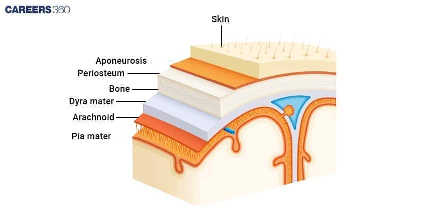
The three layers, that is, the dura mater, arachnoid mater, and pia mater, cover the brain and the spinal cord, the latter being one of the main components of the central nervous system. These layers provide support and protection for neural tissues in the brain and spinal cord.
They play an important role in maintaining the structural integrity of the central nervous system, preventing injuries to the underlying tissues and allowing for the flow of cerebrospinal fluid, which provides a "cushioning" effect to the brain and the spinal cord. By enclosing such important organs as the aforementioned ones, the meninges therein ensure proper functioning and protection from external trauma.
Brain Meninges
There are three layers in the meninges of the brain, each with a particular structure and function, to provide complete protection and support to the brain.
Structure of Brain Meninges
Brain Meninges is made up of the following components:
Dura Mater
The outer, thickest, and toughest Connective tissue in nature.
Gives a tough protective covering to the brain. And forms dural sinuses, which drain venous blood from the brain.
Arachnoid Mater
Middle and thin like a web. It is transparent.
It performs the function of a cushioning barrier. It has a subarachnoid space filled with the cerebrospinal fluid(CSF) which fluid is what cushions the brain and thus absorbs shocks.
Pia Mater
The innermost thin layer conforms to the surface of the brain.
The blood-brain barrier surrounds and protects the brain. It also cares for the blood vessels that supply the brain with nutrients and oxygen.
Blood-Brain Barrier
It is made of the endothelial cells of the brain capillaries, the surrounding astrocytes, and pericytes.
The blood-brain barrier is selectively permeable, thus providing control over the entry of substances into the brain from the bloodstream, allowing the passage of essential nutrients, but at the same time, protecting the brain from toxins and pathogens.
Spinal Cord Meninges
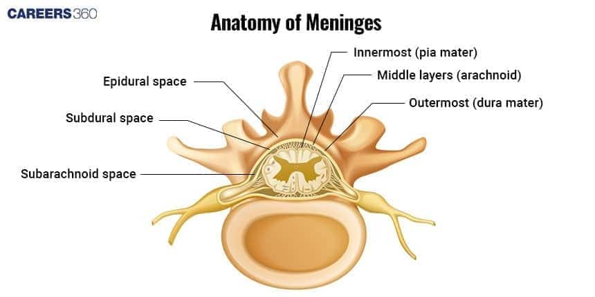
The three meninges of the spinal cord There are three layers of the meninges around the spinal cord. They provide support for the thick and long spinal cord, protect it from any shock or injury, and circulate the cerebrospinal fluid around the spinal cord.
Structure of Spinal Cord Meninges
The structure of the spinal cord is composed of:
Dura mater
The most superficial, thick, tough, and strong layer is made up of dense irregular connective tissue.
Acts as a dense, protective connector for the spinal cord to safeguard it from mechanical injury and helps stabilize the spinal cord lengthwise within the vertebral column.
Arachnoid mater
The middle layer, or the 'webby" layer, is thin and avascular.
Offers protection and contains the subarachnoid space, containing cerebrospinal fluid (CSF), which acts to cushion shocks and provide a liquid cushion to the spinal cord.
Pia mater
Pia mater is the thinnest, innermost layer that adheres to the surface of the spinal cord.
Encloses and protects the spinal cord. It contains arteries that supply nutrients and oxygen to the spinal cord tissue, and it anchors the spinal cord to the surrounding structures through the denticulate ligaments.
Protective Mechanisms
The meninges working in combination with the cerebrospinal fluid mean a multilayer protective system for guarding the spinal cord against injury and allowing the transmission of nerve impulses as well as receipt of nerve impulses without any mechanical disturbances.
Brain vs Spinal Cord Meninges — Key Differences
Difference between brain and spinal cord meninges is discussed in the table below:
| Aspect | Brain Meninges | Spinal Cord Meninges |
|---|---|---|
Thickness of Dura Mater | Thicker and more robust | Thinner and more flexible |
Attachment Points | Firmly attached to the skull | Attached to the vertebral canal via the epidural space |
Extensions | Dural folds (falx cerebri, tentorium cerebelli) extend into brain fissures | No dural folds; smooth covering |
Role in CSF Circulation | Contains cisterns that collect CSF | Circulates CSF through subarachnoid space, no large cisterns |
Protective Mechanisms | Provides rigid protection due to attachment to the skull | Offers flexibility to accommodate movement of the vertebral column |
Continuity | Continuous from the brain to the spinal cord | Continuous from the brain to the spinal cord |
Layers and Basic Composition | Three layers: dura mater, arachnoid mater, pia mater | Three layers: dura mater, arachnoid mater, pia mater |
Protection | Protects the brain from mechanical injury | Protects spinal cord from mechanical injury |
Support in CSF Circulation | Supports CSF circulation around the brain | Supports CSF circulation around the spinal cord |
Brain vs Spinal Cord Meninges NEET MCQs (With Answers & Explanations)
Important questions asked in NEET from this topic are:
Anatomy of brain and spinal cord meninges
Brain vs Spinal cord meninges
Practice Questions for NEET
Q1. The spinal cord extends from the foramen magnum at the base of the skull to the L1/L2 vertebra where it terminates as the
Conus medullaris (medullary cone)
Coccygeal vertebra
Epiduramater
Choroid plexus
Correct answer: 1) Conus medullaris (medullary cone)
Explanation:
The spinal cord, an essential component of the central nervous system, originates from the foramen magnum, a significant aperture situated at the skull's base, and extends down to the L1/L2 vertebral levels. It does not stretch throughout the vertebral column's entirety, but rather concludes at this point, transforming into the conus medullaris. Following the conus medullaris, the cauda equina, a collection of spinal nerve fibres, descends within the vertebral canal.
Hence, the correct answer is option 1. Conus medullaris (medullary cone).
Q2. The number of pairs of thoracic spinal nerves is
31
12
5
8
Correct answer: 2) 12
Explanation:
There are 8 pairs of cervical, 12 thoracic, 5 lumbar, 5 sacral, and 1 coccygeal pair of spinal nerves (a total of 31 pairs).
Hence, the correct answer is option 2) 12.
Q3. Identify the following statements.
1) The neural canal is the channel for the spinal cord to pass through.
2) The gray matter extends from the brain to the inside of the spinal cord and white matter covers the surroundings of the spinal cord.
Statement 1 is false.
Statement 2 is true.
Statements 1 and 2 are true
Statements 1 and 2 are false.
Correct answer: 3) Statements 1 and 2 are true
Explanation:
Both are true of the spinal cord:
The spinal cord is a long, cylindrical structure running from the back of the brain down to the lower part of the back and through the neural canal. It is an extremely important linkage between the central and peripheral nervous system.
The sensory neurons transmit impulses through the spinal cord. It is the portion that contains grey matter as horn-like projections and white matter surrounding it. Grey matter includes the cell bodies of neurons while white matter consists of myelinated axons connecting to other parts of the nervous system.
The spinal cord carries out the reception of sensory input and coordination of reflexes.
Hence the correct answer is option 3) Statements 1 and 2 are true.
Also Read:
Frequently Asked Questions (FAQs)
Meninges are significant for the reabsorption and circulation of CSF, especially arachnoid mater, in providing cushion to the brain and spinal cord.
Structural differences would include differences in thickness and attachment points; functional differences are related to cerebrospinal fluid dynamics
Terrible conditions can be caused by meningitis, subdural hematoma, and herniation syndromes, all of which can cause symptoms that are major and may be life-threatening, and need medical treatment.
It is an additional weight to carry due to the protection and support, for the reason that the brain is far more complex and sensitive compared to the spinal cord.
The meninges protect the central nervous system, cerebrospinal fluid circulation, and sold support provision.
