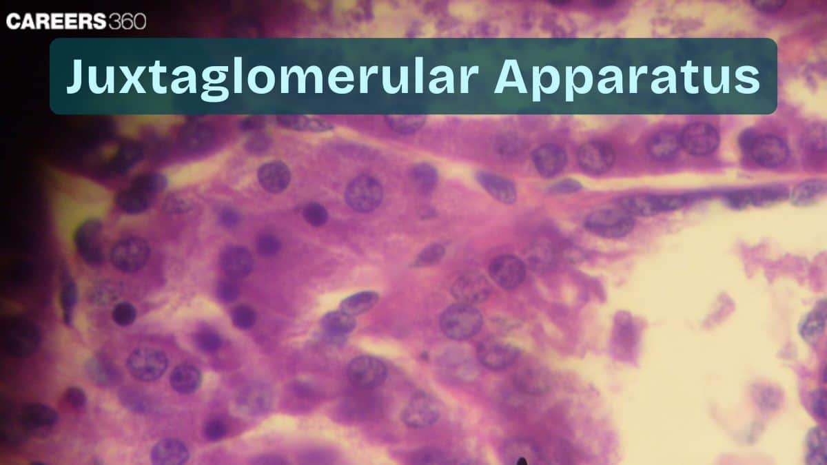
Juxtaglomerular Apparatus: Overview, Structure, Function
The Juxtaglomerular Apparatus (JGA) is a specialized region of the nephron that regulates blood pressure and glomerular filtration rate (GFR). It contains juxtaglomerular cells, macula densa, and extraglomerular mesangial cells that sense blood pressure & sodium levels and release renin. This guide covers JGA structure, functions, RAAS activation, renin secretion, GFR regulation, diagrams, FAQs, and NEET MCQs.
This Story also Contains
- What Is the Juxtaglomerular Apparatus? (Definition & Location)
- Anatomy Of The Juxtaglomerular Apparatus
- Functions Of The Juxtaglomerular Apparatus
- Juxtaglomerular Cells
- Juxtaglomerular Apparatus NEET MCQs (With Answers & Explanations)
- Recommended Video on "Juxtaglomerular Apparatus"

What Is the Juxtaglomerular Apparatus? (Definition & Location)
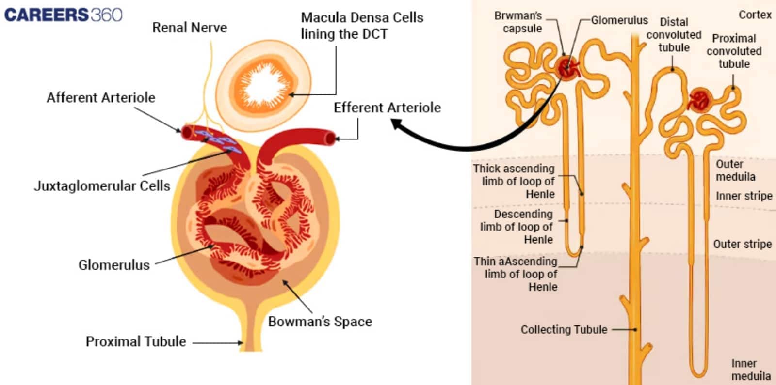
The Juxtaglomerular Apparatus is a part of the kidneys that controls blood pressure and the filtration rate by sensing changes in sodium levels through the release of renin. Located bilaterally at the junction of the afferent arteriole to the glomerulus with the distal convoluted tubule, the juxtaglomerular apparatus includes cells that have a most significant influence on regulating blood pressure and hence the filtration rate of the glomerulus. The constituent elements of the JGA are juxtaglomerular cells, macula densa cells, and extraglomerular mesangial cells.
The former monitors blood pressure and the latter monitors the sodium concentration in the filtrate. So, it secretes renin in response to low/lost blood pressure or low Na+ in the filtrate and initiates the renin-angiotensin-aldosterone system, a hormone system involved in vasoconstriction and promoting sodium and water reabsorption. This mechanism helps in maintaining proper blood pressure, sufficient for filtration and renal functioning; therefore, the JGA is essential for homeostasis.
Anatomy Of The Juxtaglomerular Apparatus
The juxtaglomerular apparatus (JGA) is located at the interface of the distal convolution, DCT, of the nephron as it curves back up to form a proximity with the afferent arteriole which feeds blood into the glomerulus.
Juxtaglomerular (JG) Cells
These are the smooth muscle cells that line the walls of an afferent arteriole. They synthesize, store, and release renin due to stimulation by low blood pressure or decreased concentration of sodium chloride.
Macula Densa
A cluster of tall, tightly fitted cells in the convoluted tubule at its distal end that come in contact with the afferent arteriole.
They detect the levels of sodium chloride in the filtrate and transmit signals for the juxtaglomerular cells to secrete renin in case of low levels of sodium.
Extraglomerular Mesangial Cells (Lacis cells)
These cells are located between the afferent arterioles and the efferent arteriole and between the glomerular capillaries.
They offer structural support, produce the extracellular matrix and transmit signals from the macula densa cells for the secretion of renin from the juxtaglomerular cells.

Functions Of The Juxtaglomerular Apparatus
The functions of the juxtaglomerular apparatus are:
Regulation Of Blood Pressure
The JGA does the important job of blood pressure homeostasis by sensing blood pressure and Na+ concentration in both blood and filtrate and bringing into action some compensatory responses in the body to rectify this disturbance.
Secretion Of Renin
The juxtaglomerular cells of the JGA are responsible for secreting the enzyme renin that converts angiotensinogen to angiotensin I—the first step in blood pressure regulation.
Role In The Renin-Angiotensin-Aldosterone System (RAAS)
The contribution of the JGA is to turn on this renin-angiotensin-aldosterone system by secreting renin. It is a system that increases the BP by vasoconstriction, and also increases the reabsorption of Na and H2O in kidneys. Hence increasing the blood volume and therefore pressure.
Maintenance Of GFR (Glomerular Filtration Rate)
The JGA changes the diameter of the afferent arteriole to maintain an optimum GFR. This would facilitate efficient filtration and prevent damage to the glomerulus due to changes in blood pressure.
Juxtaglomerular Cells
Juxtaglomerular cells are specialised smooth muscle cells located in the walls of the afferent arteriole that supply blood to the glomerulus. They lie adjacent to the convoluted tubule as it makes contact with the afferent arteriole at the distal end, forming a part of the so-called JGA.
Function In Renin Secretion
The primary role of juxtaglomerular cells is to produce, store, and secrete the enzyme renin, which plays a significant role in blood pressure regulation. In response to proper stimulation, these cells release renin into the blood where it activates the Renin-Angiotensin-Aldosterone System to raise blood pressure and thereby ensure the perfusion of the kidneys.
Stimuli For Renin Release
The juxtaglomerular cells release renin in response to several stimuli:
Low Blood Pressure: The blood pressure is locally detected by baroreceptors in the afferent arteriole.
Low Sodium Chloride Concentration: Detected by the macula densa cells in the distal convoluted tubule.
Activation of the Sympathetic Nervous System: Under stress or conditions of low blood pressure, the sympathetic nerves directly depolarize the juxtaglomerular cells.
Juxtaglomerular Apparatus NEET MCQs (With Answers & Explanations)
Important questions asked in NEET from this topic are:
Anatomy of the Juxtaglomerular Apparatus
Functions of the Juxtaglomerular Apparatus
Practice Questions for NEET
Q1. JGA is the sensitive region between
DCT and afferent arteriole
PCT and afferent arteriole
PCT and DCT
PCT, DCT and afferent arteriole
Correct answer: 1) DCT and afferent arteriole
Explanation:
The Juxta Glomerular Apparatus (JGA) is a specialized region in the kidney located between the afferent arteriole, the proximal convoluted tubule (PCT), and the distal convoluted tubule (DCT). It plays a crucial role in regulating blood pressure and the filtration rate of the glomerulus. The JGA contains specialized cells such as juxtaglomerular cells (which secrete renin) and macula densa cells (which detect sodium concentration), which work together to help maintain homeostasis by adjusting renal function in response to changes in blood pressure and sodium levels.
Hence the correct answer is option 1) DCT and afferent arteriole.
Q2. The function of macula densa is
To monitor the composition of the fluid that flows through the DCT.
To regulate renin release from the juxtaglomerular cells of the afferent arteriole.
To release the paracrine signals in the form of ATP
All of the above
Correct answer: 4) All of the above
Explanation:
The macula densa is a group of specialized cells located in the distal convoluted tubule of the nephron near the glomerulus and plays a critical role in regulating kidney function. It senses sodium chloride concentration in the tubular fluid. When NaCl levels are low, the macula densa signals the juxtaglomerular cells to release renin, which activates the renin-angiotensin-aldosterone system (RAAS) to increase blood pressure and sodium reabsorption.
Hence, the correct answer is option 4) All of the above.
Q3. A fall in glomerular filtration rate (GFR) activates
juxtaglomerular cells to release renin
adrenal cortex to release aldosterone
adrenal medulla to release adrenaline
posterior pituitary to release vasopressin.
Correct answer: 1) juxtaglomerular cells to release renin
Explanation:
The JGA plays a complex regulatory role. A fall in glomerular blood flow/glomerular blood pressure/GFR can activate the JG cells to release renin which converts angiotensinogen in blood to angiotensin I and further to angiotensin II. Angiotensin II, being a powerful vasoconstrictor, increases glomerular blood pressure and thereby GFR.
Hence, the correct answer is option 1) juxtaglomerular cells to release renin.
Also Read:
Recommended Video on "Juxtaglomerular Apparatus"
Frequently Asked Questions (FAQs)
The JGA has two key functions: it regulates the blood pressure and maintains the glomerular filtration rate and, sodium chloride, by releasing renin, which is responsible for the activation of RAAS.
The secretions of the JGA regulate blood pressure by secreting renin at low blood pressure or with a drop sodium chloride levels and hence activate the RAAS, which cause vasoconstriction and an increase in reabsorption of sodium and water, thereby increasing the blood pressure.
Low blood pressure is detected by stretch receptors in the wall of the afferent arteriole, a low concentration of sodium chloride, which is sensed by macula densa cells, and the activation of the sympathetic nervous system.
The macula densa itself senses the sodium chloride concentration in the tubule of the distal convoluted tubule and sends the signal across to the juxtaglomerular cells to release renin upon the fall of sodium levels, thus participating in blood pressure homeostasis and GFR regulation.
It is composed of juxtaglomerular cells, specialised smooth muscle cells in the afferent arteriole, macula densa cells in the distal convoluted tubule, and extraglomerular mesangial cells between the afferent and efferent arterioles.
