Mechanism of Breathing: Definition, Diagram, Functions
Breathing, or respiration, is the process of taking oxygen in and carbon dioxide. The mechanism of breathing contains two phases which are inhalation (inspiration), whereby the diaphragm contracts and the chest cavity expands, admitting air to the lungs and exhalation (expiration), whereby the diaphragm relaxes, forcing air out. The respiratory system coordinates this process for gas exchange. The understanding of the mechanism of respiration, especially in classes 10 and 11, helps explain how inspiration and expiration maintain the body's oxygen-carbon dioxide balance. This important topic from the Breathing Exchange of gases Chapter from Biology.
Don't Miss: Most scoring concepts for NEET | NEET papers with solutions
NEET 2025: Syllabus | PYQs | Crack NEET in 2 months - Study Plan
NEET Important PYQ & Solutions: Physics | Chemistry | Biology | NEET PYQ's (2015-24)
- What is Breathing?
- Process of Respiration
- Mechanism of Breathing
- Mechanism of Respiration
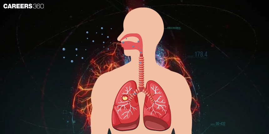
What is Breathing?
Respiration is the physiological exercise through which gases, mainly oxygen and carbon dioxide are exhaled through the lungs of an organism. This is important for cellular respiration as this moves the oxygen required in aerobic respiration to reach the cells and at the same time takes out carbon dioxide, a waste product. Proper functioning of the lungs guarantees a constant supply of oxygen to the cells for their metabolism and regulates the proper distribution of gases, which are significant for the life process.
Also Read:
Process of Respiration
The process of breathing involves the following steps:
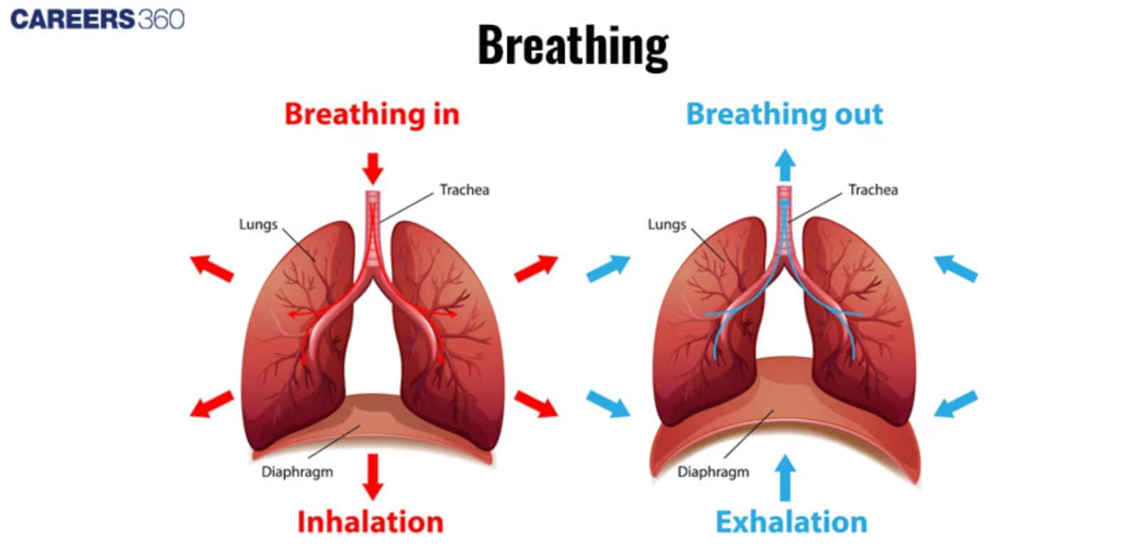
Inhalation (Inspiration)
Inhalation is mainly characterised by the contraction of the diaphragm and the intercostal muscles to raise the ribs and expand the thoracic cavity.
This contraction generates a vacuum, or negative pressure within the thoracic cavity and air rushes into the lungs.
During the process of inhalation, contraction of the muscles surrounding the thoracic cavity increases the volume of this cavity and thus forces the air through the respiratory tract and into the alveoli to participate in the exchange of gases.
Exhalation (Expiration)
Exhalation takes place when the diaphragm and intercostals decrease their size decreasing the volume of the thoracic cavity.
This relaxation leads to positive pressure in the thoracic cavity which in turn forces air out of the lungs and out of the respiratory tract.
The reduction of volume ejects the air laden with carbon dioxide ending the breathing cycle.
Mechanism of Breathing
The mechanism of breathing involves the following:
Diaphragm and Intercostal Muscles
The diaphragm functions as a muscle which is in the shape of a dome and is situated at the bottom part of the thoracic cavity and the intercostal muscles are located in between the ribs of an individual’s body.
Inhalation is done by the contraction of the diaphragm, which becomes flattened and the intercostal muscles that lift the ribs, expanding the thoracic cavity. By doing so, the pressure in the thoracic cavity is reduced enabling air to enter the lungs.
These muscles contract during inhalation and, at the same time, they relax during exhalation to narrow the thoracic cavity hence forcing air out of the lungs.
Lung Compliance and Elasticity
Lung compliance on the other hand deals with the ease with which the lungs can expand and dilate given some pressure.
High lung compliance means that the lungs can be expanded easily and can therefore fill up easily while low compliance means that the lungs are stiff and cannot be filled easily.
Accessibility of the lungs can be affected by the degree of elasticity of the lung tissues, and the quantity and nature of the surface tension-reducing agents. This leads to disorders in respiration.
Lung compliance is an index of work that the respiratory muscles need to do to achieve a certain level of ventilation.
Airway Resistance
Airway resistance is the opposition force within the respiratory tracts to the flow through them.
This depends on the dimensions like the diameter of the airway, mucus production, and bronchial constriction.
There is always the case of high resistance, this is because of the constriction of the tubes through which air has to pass, or the presence of an obstructing factor that makes it difficult to breathe.
It is important to deal with airway resistance if breathing properly and if fresh and expired air is going to get to the lungs.
Mechanism of Respiration
Some important mechanisms of respiration are discussed below:
Intrapleural Breathing
The pressure in the pleural cavity, the space between the lungs and pleura, is referred to as intrapleural breathing. This pressure is less than that of atmospheric pressure, known as negative pressure, which is very vital in the mechanics of respiration. The transpulmonary pressure or the pressure difference is accountable for the lung movement during breathing. The intrapleural pressure becomes more negative during inhalation and causes the lungs to expand. During expiration, the pressure increases, causing the lungs to recoil.
The lungs' elasticity and the surface tension of alveolar fluid pull the lungs inward, while the thoracic wall and pleural fluid create an opposing force. This balance results in negative intrapleural pressure, a key concept in the mechanism of breathing.
Respiratory Gas Transport
Respiratory gas transport refers to the transport of oxygen and carbon dioxide by the blood. Oxygen-rich blood from the lungs flows to the heart through pulmonary veins. The heart pumps this oxygenated blood to the rest of the body through the aorta.At the same time, deoxygenated blood with carbon dioxide is returned to the lungs by the pulmonary arteries for gas exchange. The whole process is repeated continuously so that oxygen, carbon dioxide, and carbon dioxide balance are maintained in the body.
This explanation of the mechanism of breathing, how to inspire and expire, helps to describe more precisely how the respiratory system works in a human being.
The gas exchange in the lungs involves the following:
Diffusion of Gases
In the alveoli oxygen from the inhaled air moves passively through the walls of the alveoli into the blood in capillaries, and at the same time, carbon dioxide diffuses active from the blood into the alveoli and thus is exhaled.
This exchange is driven by partial pressure gradients. Oxygen diffuses from the place that has high partial pressure in the alveoli relative to that found in blood and similarly for the diffusion of carbon dioxide.
Hemoglobin and Oxygen Transport
Oxygen on reaching the lungs gets attached to the haemoglobin molecules and forms oxyhemoglobin which is easily transported to the tissues.
The oxygen dissociation curve shows how the affinity of haemoglobin for oxygen varies with different concentrations of oxygen partial pressure.
At high partial pressures, for example in the lungs, haemoglobin grabs more oxygen than from the lower pressure found in tissues where oxygen is needed, for use by the cells.
Also Read
Recommended video for "Mechanism of Breathing"
Frequently Asked Questions (FAQs)
Respiration is the process by which gases are taken into the lungs and expelled out of the lungs through the act of breathing. When the diaphragm and other such as intercostal muscles contract and expand the thoracic cavity the pressure drops and air rushes into the lungs. Expiration takes place when these muscles relax reducing the volume of the thoracic cavity and applying pressure on the lungs to force out the air.
The diaphragm is a conical muscle which splits the thoracic cavity from the abdominal cavity. When the person breathes in, it relaxes and becomes thin to expand the volume of the thoracic cavity and produce a negative pressure that sucks in air into the lungs. When breathing out, the diaphragm becomes flattens, hence decreasing the volume of the thoracic cavity and forcing an expulsion of air from the lungs.
Inspiration is the process of taking in air into the lungs and is accompanied by contraction of the diaphragm and the intercostal muscles which causes a negative pressure in the thoracic cavity.
Expiration is the process of the removal of air from the lungs and is characterized by relaxation of the diaphragm and intercostal muscles, hence leading to the reduction of the thoracic cavity volume and the pressure increases leading to expulsion of air.
This is through the respiratory centre in the brainstem, especially the medulla oblongata and pons. These centres check the levels of the concentration of carbon dioxide, amount of oxygen and blood pH and modify the depth and frequency of breath. Other specialists also called sense organs help to feedback to various organs particularly the respiratory centre to ensure proper gas exchange and stability.
Some of the diseases affecting the respiratory system are asthma, which involves inflammation and constriction of the airways; COPD embracing emphysema and chronic bronchitis cause obstruction of the airways; pneumonia, which is an infection affecting the lungs; and pulmonary fibrosis, which results from the formation of scar tissues in the lungs hence restricting breathing.
Also Read
29 Nov'24 11:00 PM
27 Nov'24 01:11 PM
26 Nov'24 11:17 AM
26 Nov'24 10:17 AM
25 Nov'24 07:51 PM
25 Nov'24 07:19 PM
25 Nov'24 11:48 AM
18 Sep'24 06:39 PM
18 Sep'24 06:35 PM
18 Sep'24 04:11 PM