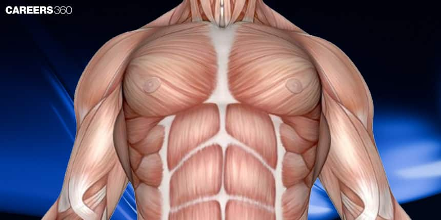
Muscle Contraction And Contractile Proteins: Definition, Explanation, Function
Muscle contraction is the process that enables movement in living organisms by shortening and generating tension within muscle fibres. It occurs through a coordinated interaction between contractile proteins, primarily actin and myosin, along with troponin and tropomyosin. These proteins work together in response to signals from the nervous system, using energy from ATP. In this article, muscle contraction, contractile proteins, the anatomy of muscle tissue, types of contractile proteins, and the mechanism of muscle contraction are discussed. Muscle contraction and contractile proteins are a topic of the chapter Locomotion And Movement in Biology.
- Definition of Muscle Contraction
- What are Contractile Proteins?
- Anatomy of Muscle Tissue
- Types of Contractile Proteins
- The Mechanism of Muscle Contraction

Definition of Muscle Contraction
Muscle contraction is a process whereby muscle fibres develop tension and obtain shortening or lengthening to enable movement and generation of forces through the body. It is a foremost vital process in various biological functions, starting from voluntary movements like locomotion to continuing posture producing heat, and protecting viscera. This means that the said organ takes part in the performance and functioning of the heart and the process of moving substances by the digestive and vascular systems.
This article consists of extensive information regarding muscle contraction, whereby it describes, with detail, how muscle contraction works, describing their involvement right from the neuromuscular junction, the excitation-contraction coupling, and the cross-bridge cycle. It also explains the multiple muscle fibres and diverse sources of energy that fuel the muscle contractions, as they pertain to being significant for health and human bodily full operation as well
What are Contractile Proteins?
Contractile proteins are a special kind of protein witnessed in muscle cells. Two key contractile proteins, actin and myosin, are very vital in muscle contractility and movement. Actin is a thin filament, and myosin is a thick filament with little projections binding to actin to cause force. During muscle contraction, myosin heads form a bond with the actin filament and swing in what is referred to as a power stroke or stroke of energy that drags the actin filaments in, consequently shortening the muscle fibre. This interaction is controlled by other proteins, such as troponin and tropomyosin, which regulate the bond of actin and myosin in response to the concentration of calcium ions.
Also Read:
- MCQ Practice for Muscle Contraction and Contractile Proteins
- Locomotion and Movement
- Human Skeletal System
- Joints
Anatomy of Muscle Tissue
The anatomy of muscle tissue is discussed below-
Types of Muscle Tissue
The different types of muscle tissue are:
Skeletal Muscle
It is striated. It comes under voluntary control. It is attached to the bones using the tendons and so is in charge of movement and posture. The muscle fibres are long, cylindrical cells with multiple nuclei peripherally placed.
Cardiac Muscle
Present only in the heart, cardiac muscle is striated and involuntary. It is composed of very short cells, branching and interconnected to other cells by intercalated discs. The presence of these discs enables the cells to contract in a coordinated fashion, responsible for the pumping action of the heart.
Smooth Muscle
In this class of muscles, one does not find striations, and functioning is unconscious. It exists lining the inner walls of internal organs, like the digestive tract and blood vessels. The muscle fibres usually have a spindle shape with a single centrally located nucleus and carry out movements like peristalsis and vasoconstriction.
Structure of Skeletal Muscle
The structure of muscle fibre includes:
Muscle Fibre
The fibre is the single cellular unit of skeletal muscle tissue. It presents as a long structure with a cylindrical shape. It is enveloped by a plasma membrane, referred to as a sarcolemma. Each fibre is multinucleated and contains many myofibrils, and each is capable of contraction.
Myofibrils
These are individual subunits of the muscle fibre, where the contractile proteins—actin and myosin—are housed. Normally, they happen to line up parallel to each other and in orientation within the fibre. They are grouped into units called sarcomeres, which are the functioning units for contraction in muscles.
Sarcomere
A sarcomere is the simplest contractual entity of a muscle fibre, bounded on both ends by Z-lines. It overlaps the thick (myosin) and thin (actin) filaments of the muscle fibres, whose interactions bring about muscle contraction—shortening of the sarcomere.
Types of Contractile Proteins
The contractile proteins are discussed below-
Actin
Actin is a globular protein that polymerises to form thin filaments in muscle fibres; each actin filament is a double helix of actin subunits with sites available for binding with myosin heads. The actin filaments provide the track along which the myosin heads move during muscle contraction.
Interaction with Myosin
The actin and myosin interact through the process of muscle contraction by making cross-bridges. For this to happen, myosin becomes attached to pinpoint bindings on the actin filament so that the myosin can drag the actin filaments inwards resulting in the shortening of the sarcomere and thus in muscle contraction.
Myosin
One of the thick filament proteins, myosin is typical, having a long, fibrous tail and a globular head. Into the head portion, myosin molecules possess ATPase activity, which is very important during the production of energy for contraction. Its primary function is the interaction with actin to create force and motion.
Myosin Head and Power Stroke
The myosin head binds to the actin molecule to form a cross-bridge. During the power stroke, the myosin head swivels, which pulls the actin filament toward the centre of the sarcomere. This results in the shortening of the sarcomere and the creation of tension within the muscle. Once ATP is attached to the myosin, the myosin head releases from the actin and reattaches to cock for the next cycle.
Tropomyosin And Troponin
Regulation of Muscle Contraction
Tropomyosin and troponin are proteins that act as regulators for the action of actin with myosin. In resting muscle, tropomyosin masks the sites on the actin filament to which myosin does not bind. Muscle contraction occurs because of a conformational change in the troponin as a result of the binding of tropomyosin to calcium ions that move tropomyosin out of blocking those sites.
Interaction with Actin and Myosin
Attached to the actin filament is tropomyosin, and attached to tropomyosin is troponin. Calcium binding to troponin causes a conformation change; this change causes movement in tropomyosin quickly enough to expose sites where myosin will attach to the actin molecule. There myosin heads attach to the actin, and contraction begins.
The Mechanism of Muscle Contraction
The mechanism of muscle contraction is discussed below-
Sliding Filament Theory
Thus, in the sarcomere, this happens through the sliding motion of the actin and myosin filaments. During contraction, myosin heads bind to the actin and pull it toward the centre of the sarcomere using the power stroke.
This results in the shortening of the sarcomere, which leads to a contraction of the muscular tissue. Here, the theory extends to the dynamic aspect of the movement of filaments, whereby the constant filament length changes in their overlap lead to short muscles.
Role of Calcium Ions
Calcium ions usually play a very fundamental role in the muscle contraction process, since they orchestrate in the actin-myosin interaction. After a muscle fibre has been stimulated, calcium ions flow from the sarcoplasmic reticulum into the sarcoplasm.
Calcium is bound to troponin, which creates a conformational change that rolls the tropomyosin away from myosin-binding sites on actin. This exposure finally makes myosin heads possible to bind and initiate contraction. Removal of the calcium ions stops the reaction, and thus the muscle will finally relax.
ATP and Muscle Contraction
ATP is the energy source that drives muscle contraction. It does this by energizing the myosin head, so that it may bind to actin, perform the power stroke of contraction, and then be unbound from the actin filament.
It is also used in the recocking of the myosin heads and in the active transport mechanisms that pump Ca2+ back into the sarcoplasmic reticulum after a contraction has ceased. If ATP were not continuously available, the muscle would pause in its activity, since its contraction and recovery require energy obtained only through the hydrolysis of ATP.
The Neuromuscular Junction
Synapse
The NMJ is a specialised synapse by which a motor neuron communicates with a muscle fibre; thus, the NMJ consists of the axon terminal of the motor neuron, the synaptic cleft, and the motor end plate on the muscle fibre. The junction serves to transmit the nerve impulse for pertaining muscle contraction.
Neurotransmitters
The neurotransmitter at the NMJ, from the motor neuron, is acetylcholine. The ACh binds onto the motor end plate of the muscle fibre and influxes sodium ions, generating an action potential. The action potential is propagated along the muscle fibre. The action potential results in muscle contraction through the release of calcium ions from the sarcoplasmic reticulum.
Also Read:
| Ribs and Rib Cage | Vertebral Column |
| List of 206 Bones in Body | Pectoral Girdle |
| Axial Skeleton System | Appendicular Skeleton System |
Recommended video on Muscle Contraction
Frequently Asked Questions (FAQs)
Muscle contraction is the process during which muscle fibres develop tension and eventually shorten. What will the myosin heads bind to and how during this process of muscle contraction do the actin filaments get pulled in toward the centre of the sarcomere to shorten?
The major contractile proteins are actin and myosin. Actin makes up the thin filaments, and myosin makes up the thick filaments. A myosin head attaches to the actin and pulls towards itself, which incurs muscle contraction.
This theory says that the filaments slide against each other, thus causing the contraction of a muscle. Myosin heads bind with actin, pull it inward, and let go, and the result is a shortening of the sarcomere.
From the question above, calcium ions bind to troponin and that leads to shifting of tropomyosin off myosin binding sites on actin, which is then allowed to be exposed so that myosin heads bind and initiate contraction.
The different kinds of muscle contractions are isotonic: muscle length does change.
Also Read
02 Jul'25 06:47 PM
02 Jul'25 06:47 PM
02 Jul'25 06:47 PM
02 Jul'25 06:46 PM
02 Jul'25 06:46 PM
02 Jul'25 06:46 PM
02 Jul'25 06:46 PM