

Nephron- Function Of Renal Tubules: Definition, Structure, Diagram, & Facts
Nephrons are the structural and functional units of the kidney responsible for filtration, reabsorption, secretion, and urine formation. Each nephron contains a renal corpuscle and renal tubule—PCT, Loop of Henle, DCT, and collecting duct—working together to maintain fluid, pH, and electrolyte balance. This guide covers nephron structure, functions of each tubule segment, counter-current mechanism, hormonal control, diagrams, FAQs, and NEET MCQs.
This Story also Contains
- What is Nephron?
- Structure of a Nephron
- Functions of Renal Tubules
- Nephron NEET MCQs (With Answers & Explanations)
- Recommended Video for "Nephron- Function Of Renal Tubules"
What is Nephron?
The nephron is the structural and functional unit of the kidney and is, thus, involved in an important role concerning blood filtration and homeostasis, much like the entire renal system. There are around one million nephrons in each kidney that remove waste products, balance electrolytes, and regulate blood pressure.
The nephron is composed of a renal corpuscle and renal tubules, which further comprise a proximal convoluted tubule, a Loop of Henle, a distal convoluted tubule, and a collecting duct. All these parts work as a group to carry out the vital processes of filtration, reabsorption, secretion, and excretion to maintain homeostasis and ensure a body remains fit and healthy.
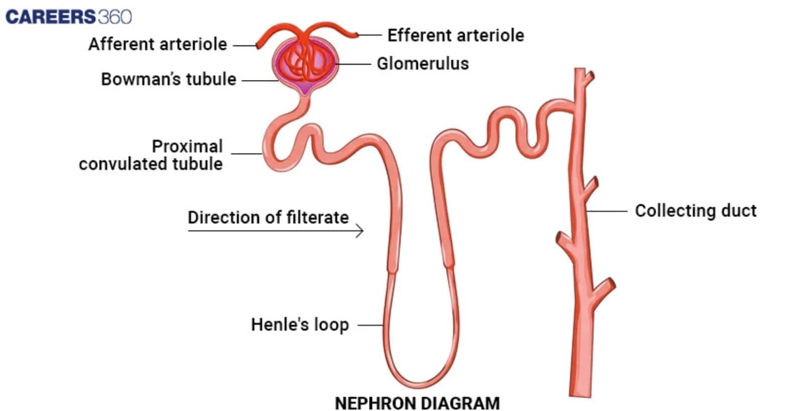
Structure of a Nephron
The structure of the nephron is discussed below-
Renal Corpuscle
Glomerulus: The glomerulus sits at the very start of the nephron and conducts the initial step of filtering the blood. As a result of the severe blood pressure the water, salts and small molecules are forced out from the blood into Bowman's capsule, thus producing the filtrate.
Bowman's Capsule: The Bowman's capsule is a double-walled cup-like structure. It encloses the glomerulus and receives the filtrate secreted from the glomerulus, which it conveys to the renal tubules to continue with the process. Renal Tubules
Renal Tubules
Proximal Convoluted Tubule (PCT): This is the first part of the renal tubule which comes out from Bowman's capsule. The PCT is highly coiled, thus providing a large surface area for reabsorption with the lining of microvilli. Significant reabsorption of water, glucose, ions, and other vital nutrients into the bloodstream occurs here.
Loop of Henle: In this region, one would expect to see the loop of an arch extending into the renal medulla. This would contain the descending limb responsible for re-absorbing water and the ascending limb, which re-absorbs salts. The Loop of Henle is involved in an essential process in the concentration of urine.
Distal Convoluted Tubule (DCT): More reabsorption and secretion take place here. Under the control of hormones, the DCT fine-tunes filtrate composition with the reabsorption of more ions and water, e.g., aldosterone.
Collecting Duct: It receives urine from many nephrons and further processes it to end the process of urine concentration by the reabsorption of water under the control of ADH. Concentrated urine is then transported towards the renal pelvis and then into the ureter.
Functions of Renal Tubules
The function of the nephron is discussed below-
PCT Functions
Reabsorption at the PCT comprises mainly essential nutrients, such as glucose, and amino acids, and ions such as sodium, potassium, and bicarbonate.
In the PCT, approximately 65-70% of the filtrate is reabsorbed.
Some waste products, such as hydrogen ions, creatinine, and some drugs and toxins, are secreted by the PCT into the filtrate to be eliminated as urine.
Loop of Henle Functions
The descending limb of the Loop of Henle reabsorbs water since it is permeable to water though it's not permeable to salts. As the filtrate descends, water moves out of the filtrate and into the surrounding hypertonic medulla, hence concentrating the filtrate.
The ascending limb is impermeable to water. It is the site where active transport expels sodium and chloride ions from the filtrate, thus decreasing the osmolarity of the filtrate.
The countercurrent multiplier system of the Loop of Henle establishes a gradient in the renal medulla. This gradient is what allows the kidney to produce concentrated urine.
DCT Functions
The DCT makes state adjustments in the amount reabsorbed based on the needs of the body.
Many ions (such as potassium, sodium, and calcium) are controlled there, along with the control of blood pH by the secretion of hydrogen versus the reabsorption of bicarbonate.
The functions of DCT are influenced by aldosterone and ADH.
For instance, aldosterone increases the re-absorption of sodium and, therefore, in case of more re-absorption of sodium, the secretion of potassium also increases in an equal amount of sodium re-absorbed.
While ADH increases the reabsorption of water.
Collecting Duct Functions
It is responsible for regulating the final concentration of the urine to be excreted. It reabsorbs water under the influence of ADH.
There is more water passage through the wall of ducts due to increased permeability.
Water and electrolyte balance gets regulated at the collecting ducts by reabsorption of sodium and secretion of potassium and hydrogen ions.
The collecting duct also contributes to the secretion of hydrogen ions and reabsorption of bicarbonate in maintaining acid-base balance.
Nephron NEET MCQs (With Answers & Explanations)
Important questions asked in NEET from this topic are:
Structure of Nephron
Functions Renal Tubule
Practice Questions for NEET
Q1. Middle protective layer seen in kidneys of human is
Renal capsule
Renal fascia
Renal peritoneum
Adipose layer
Correct answer: 4) Adipose layer
Explanation:
The layers surrounding the kidney are three.
a) Renal fascia (outermost) - This is the dense connective tissue layer that provides support and protection to the kidneys and adrenal glands.
b) Adipose capsule (intermediate) - This is known as perirenal fat; it surrounds the kidneys and serves to cushion and insulate them.
c) Renal capsule (innermost) - A thick fibrous layer that directly encases the kidney, providing further protection and maintaining its shape.
Hence the correct answer is Option (4) Adipose layer
Q2. Columns of Bertini in the kidneys of mammals are formed as extensions of
Cortex in the medulla
Cortex in pelvis
Medulla in pelvis
Pelvis in ureter
Correct answer: 1) Cortex in the medulla
Explanation:
Columns of Bertini-The renal cortex extends between the renal pyramids in the kidney to form the columns of Bertini, also referred to as renal columns. They consist of fibrous material, urine tubes, and blood arteries. The cortex is anchored in place by the Bertini columns.
Hence, the correct answer is Option (1) Cortex in the medulla.
Q3. Skin can eliminate certain substances like
Sweat
NaCl
Urea
All of these
Correct answer: 4) All of these
Explanation:
The sweat and sebaceous glands in the skin can eliminate certain substances through their secretions. The sweat produced by the sweat glands is a watery fluid containing NaCl, small amounts of urea, lactic acid, etc. This procedure aids in both thermoregulation and the body's removal of trace amounts of waste.
Hence, the correct answer is option 4)All of these.
Also Read:
Recommended Video for "Nephron- Function Of Renal Tubules"
Frequently Asked Questions (FAQs)
Water gets reabsorbed in the descending limb, thereby concentrating the filtrate; on the other hand, sodium and chloride ions are reabsorbed in the ascending limb without water to follow, thereby creating a gradient that helps concentrate urine in the medulla.
ADH (antidiuretic hormone) is released to increase water reabsorption in DCT and collecting duct. Aldosterone acts on the DCT and causes increased reabsorption of sodium and increased secretion of potassium.
ATN, PKD, and RTA are some of the more common disorders among the many that affect the renal tubules.
Among the important substances reabsorbed in the PCT are water, glucose, amino acids, sodium, potassium, bicarbonate, and other vital nutrients. This is an important stage in conserving imperative nutrients, water, and electrolytes in the body.
The major function of the renal tubules is to process the filtrate produced by the glomerulus through reabsorption, secretion, and excretion, such that the proper body balance of water, electrolytes, and waste products are maintained and urine is formed ready to be excreted out of the body.
