Parts of Human Excretory system: Organs, Functions, Diagrams
The human excretory system removes waste products and maintains fluid balance in the body. Its major components include the kidneys, ureters, urinary bladder, and the urethra. This topic is from the Class 11 chapter Excretory Products and Their Elimination of Biology. It is an important topic for competitive exams like NEET, and AIIMS BSc Nursing since questions are asked related to the structures and functions of the excretory system as well as the mechanisms involved in maintaining homeostasis.
- What is the Human Excretory System?
- Organs of the Human Excretory System
- Kidneys
- Ureters
- Urinary Bladder
- Urethra
- Mechanisms of Urine Formation
- Dialysis
- Recommended Video on Parts of Human Excretory System
What is the Human Excretory System?
The excretory system in the human body is a network of organs and structures that play a role in the removal from the bloodstream of avoidable products and extra amounts of substances, thereby ensuring balance in the components of the body. This includes the kidneys, ureters, urinary bladder, and urethra. The kidneys filter blood for urea, toxins, and excess water in the blood to form urine. The urine then passes through the ureters down to the bladder for temporary storage and excretion through the urethra. In its regulatory role over the composition of the blood, the levels of electrolytes, and the amount of fluid, it helps in the proper functioning of cells and organs, thereby maintaining proper health and stability of the body.

Also Read:
- Excretory Products and their Elimination
- Human Excretory System
- Disorders of Excretory System
- Locomotion and Movement
Organs of the Human Excretory System
The human excretory system removes waste products from the body. It has several major parts, including:
- Kidneys: These are bean-shaped organs that filter blood to remove waste, excess salts, and water, which is converted into urine.
- Ureters: Thin tubes that carry urine from each kidney to the bladder.
- Urinary Bladder: A muscular sac that stores urine until it is ready to be excreted.
- Urethra: It is a tube that conveys urine from the body outside during urination out of the bladder.
Together, these organs regulate water balance, eliminate waste products, and ensure overall homeostasis in the body.
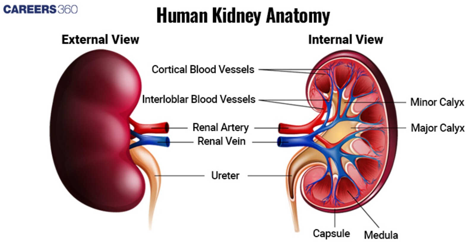
Kidneys
The kidneys are a pair of bean-shaped organs, sitting on either side of the spine between the rib cage. Each of the kidneys measures about 11-14 cm in length and 150 grams in weight.
The following three important parts differentiate in the kidney:
Renal Cortex: It is the outer layer comprising glomeruli with their convoluted tubules and hence forms the commencement site for blood filtration.
Renal Medulla: The inner region of the kidney is composed of renal pyramids. These pyramids include the loops of Henle and collecting ducts, which are significant for the concentration of urine.
Renal Pelvis: Funnel-shaped cavity collecting the urine from renal pyramids and channels into the ureter.
Diagram of the Kidney
Functions of the Kidney
The functions of the kidney are:
Filtration of blood
The kidneys filter around 180 litres of blood every day. Waste products, and excess substances, along with water, are removed from the blood in the glomeruli to result in urine.
Reabsorption and secretion
During the passage of filtrate through renal tubules, important nutrients, ions, and water are reabsorbed into the bloodstream from it. While this is in process, further waste products and excess ions get secreted into the filtrate from the blood to adjust the balance of the body perfectly.
Production of urine
The filtrate, now called urine, is collected in the renal pelvis. Through the ureters, this urine is whisked away to be stored in the bladder before being excreted out of the body. The volume and composition of urine are regulated by the kidneys to maintain electrolyte balance, pH, and overall fluid homeostasis.
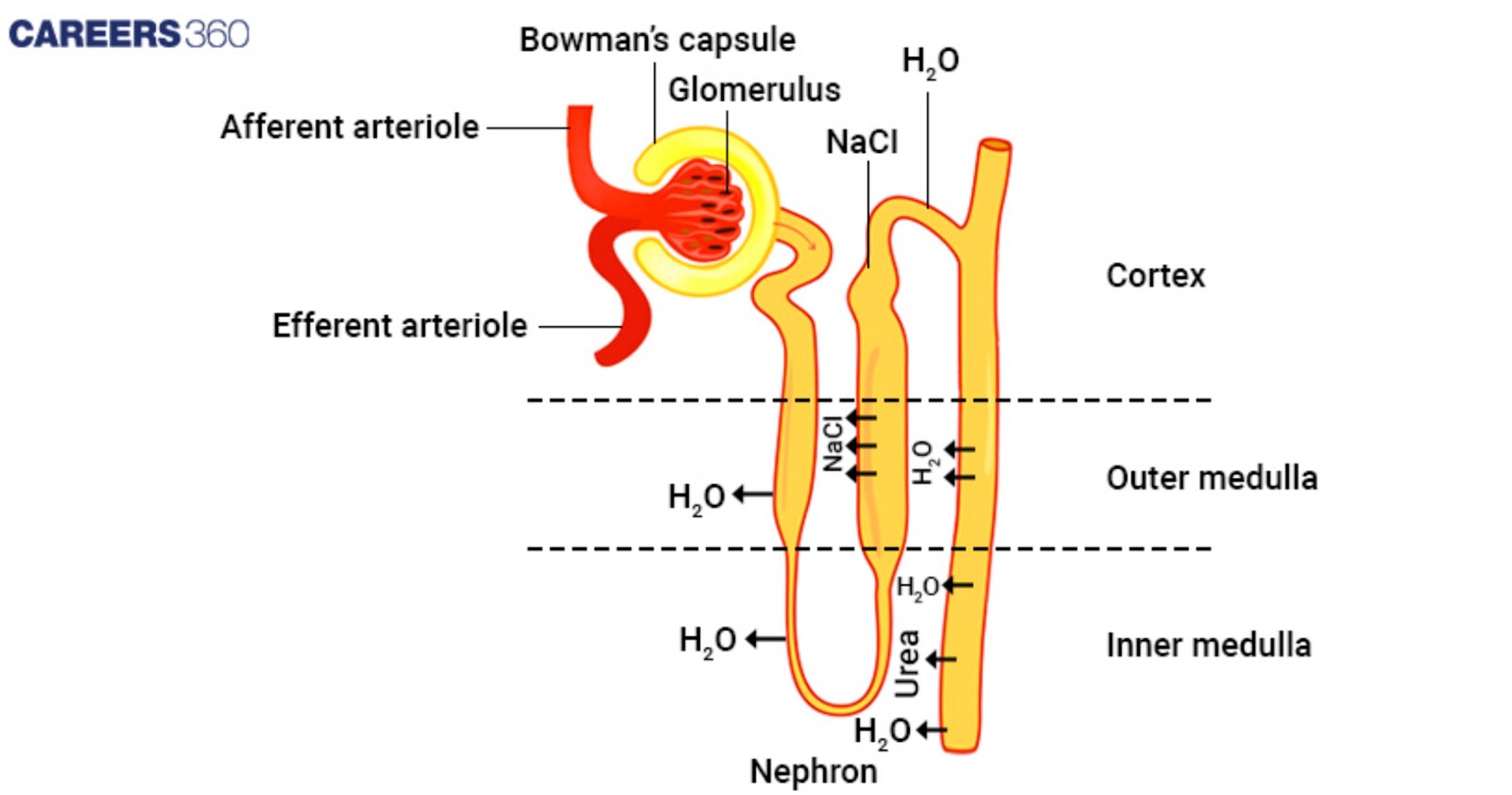
Nephron: The Functional Unit of the Kidney
The nephron is the microscopic functional unit of the kidney, and its structural counterpart is a million such units in each kidney.
Each nephron consists essentially of a renal corpuscle to which is attached a renal tubule.
Bowman's Capsule: A cup that covers the glomerulus, it acts as the site of primary blood filtration—a pressure filter—where all the water and small molecules get filtered into the renal tubule.
Glomerulus: A network of capillaries within the Bowman's capsule, responsible for filtering the blood. It allows only water, ions, and small molecules through while letting large molecules such as proteins and blood cells remain behind.
Proximal Convoluted Tubule (PCT): the part of the nephron that lies immediately distal to Bowman's capsule, where huge reabsorption of water, ions, and nutrients is done. Plus, it secretes waste products into the filtrate.
Loop of Henle: This is a U-shaped segment that extends into the renal medulla; it contains descending and ascending limbs, viewed as most important for concentrating urine by reabsorption back both water and sodium chloride.
Distal Convoluted Tubule: The part beyond the loop of Henle that calls forth selective reabsorption and secretion of ions, recognized as being very vital for maintaining correct acid-base balance and levels of various electrolytes within the body.
Collecting Duct: The last portion that collects the urine from several nephrons and discharges it via the renal medulla to the renal pelvis. It is a key site for the regulation of water reabsorption under the influence of antidiuretic hormone (ADH).
Diagram of the Nephron
Function of the Nephron
The following are the main functions of the nephron:
Filtration
As blood enters the glomerulus, it is filtered across the glomerular capillaries into Bowman's capsule, forming the glomerular filtrate. This filtrate contains water, ions, glucose, and small molecules but does not include large proteins and blood cells.
Reabsorption
While passing through the proximal convoluted tubule, the loop of Henle, and the distal convoluted tubule, useful substances such as water, glucose, amino acids, and ions are reabsorbed into the bloodstream.
Secretion
The nephron processes and secretes more waste products and extra ions from the blood into the tubular fluid. Most of the tubular secretion takes place in the proximal and distal convoluted tubules, where final adjustments in the filtrate's composition occur.
Excretion
The collecting ducts gather the final product or urine and pass it down to the renal pelvis. The urine then flows, through the ureters, into the bladder, at which point it is stored until it is excreted from the body.
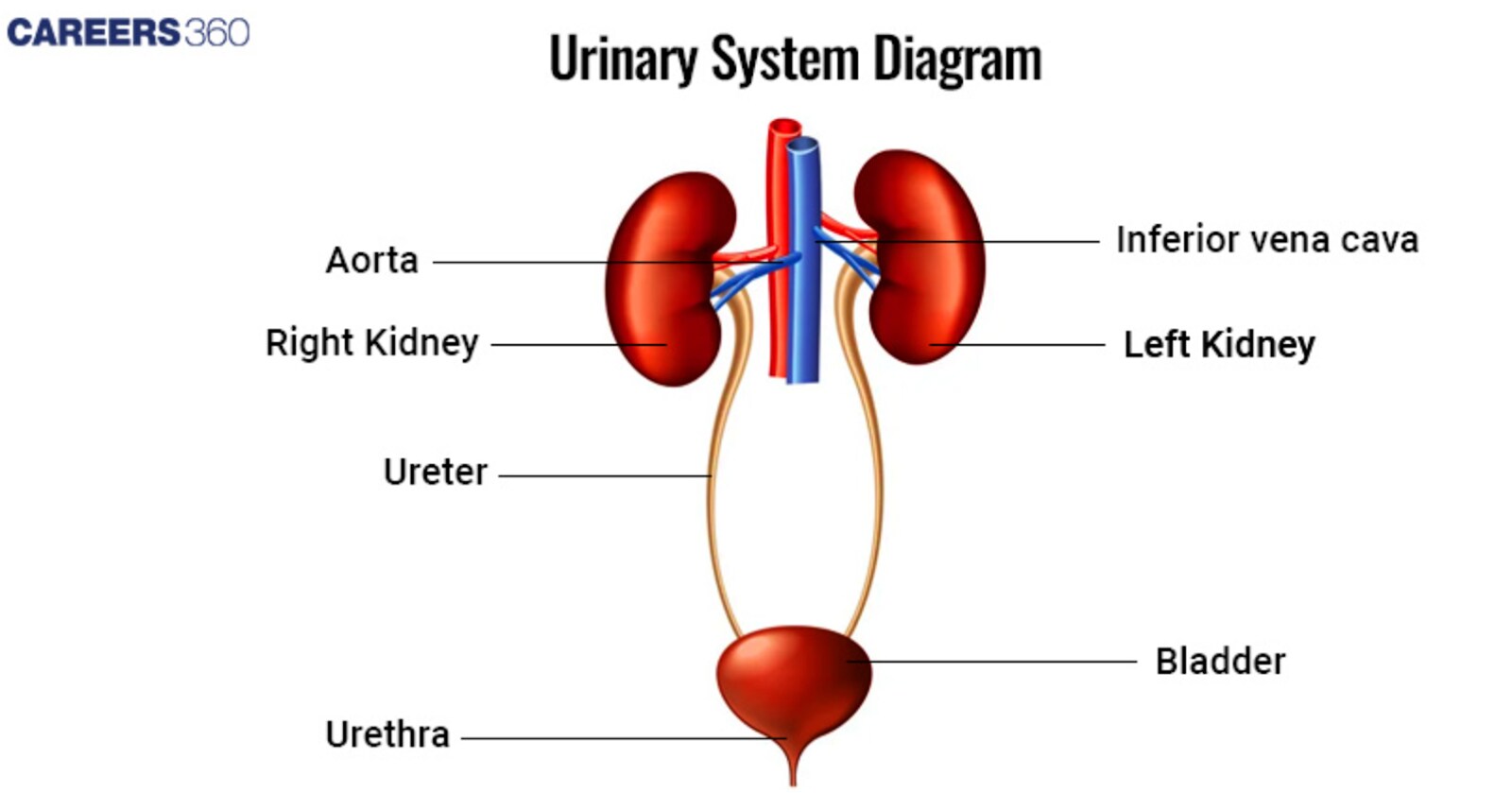
Ureters
The two slender, muscular tubes of the ureters extend from the renal pelvis of each kidney to the bladder. They are approximately 25–30 cm long and about 3–4 mm in diameter. The walls are composed of three layers: an inner mucosal layer, a middle muscular layer, and an outer adventitia layer.
- Inner Mucosal layer: This layer is made of transitional epithelium. This kind of epithelium can expand which is essential to the ureters when urine flows through them.
- Middle Muscular Layer: The middle layer is composed of smooth muscle fibres aligned circularly and longitudinally in successive layers. The successive muscular layers contract peristaltically, pushing the urine towards the bladder.
- Outer Adventitia Layer: The outermost layer is composed of fibrous connective tissue and acts as a framework for support to affix the ureters in position.
Diagram of the Ureters
Function of the Ureters
The functions of the ureters include:
Transport of urine from the kidneys to the bladder
The movement of urine from the renal pelvis of each kidney down into the bladder is accomplished by the ureters. Each ureter accomplishes this through the countercurrent smooth muscle contractions of its walls. By these repetitive contractions, urine is effectively squeezed down toward the bladder without backflow to interfere with the continual efforts of the kidneys to produce and eliminate urine.
Urinary Bladder
The urinary bladder is a sac-like muscular organ located inside the pelvis, behind the pubic bone. It is a temporary reservoir for urine. Its shape is triangular when empty and becomes more spherical as it fills with urine. The bladder's capacity in adults generally varies between 400 to 600 millilitres.
Mucosa: The innermost layer of the bladder wall, and is lined with transitional epithelium. Transitional epithelium can stretch a significant amount; therefore, it permits the bladder to stretch as it fills with urine. The mucosa also has rugae, or folds, which also allow for stretching.
Muscularis (Detrusor Muscle): This layer comprises a thick smooth muscle with fibres directed into three planes (inner longitudinal, middle circular, and outer longitudinal). This muscle is responsible for squeezing the bladder empty.
Serosa: Superficial to the muscularis is a serosa or an adventitia over areas of the urinary bladder that are not coated with the peritoneum. The serosa holds the shape and structure of the bladder.
Diagram of the Urinary Bladder

Function of Urinary Bladder
The main functions of the urinary bladder are:
Storage of urine
The auxiliary function of the urinary bladder is to store urine produced by the kidneys, where it is stored until finally being excreted. The bladder can hold quite large quantities of urine due to its expandable nature, allowing urine release to be regulated and timed in intervals.
Mechanism of urine release (micturition)
The mechanism of urination is a cooperative arrangement of involuntary and voluntary mechanisms. When the urinary bladder is filling up with urine, stretch receptors develop in the bladder wall. Impulses are sent to the brain, which initiates the general sensation of needing to micturate. At the same time, the detrusor muscle contracts, and the internal (involuntary) and external (voluntary) urethral sphincters relax, allowing the flow of urine from the bladder through the urethra into the outside world.
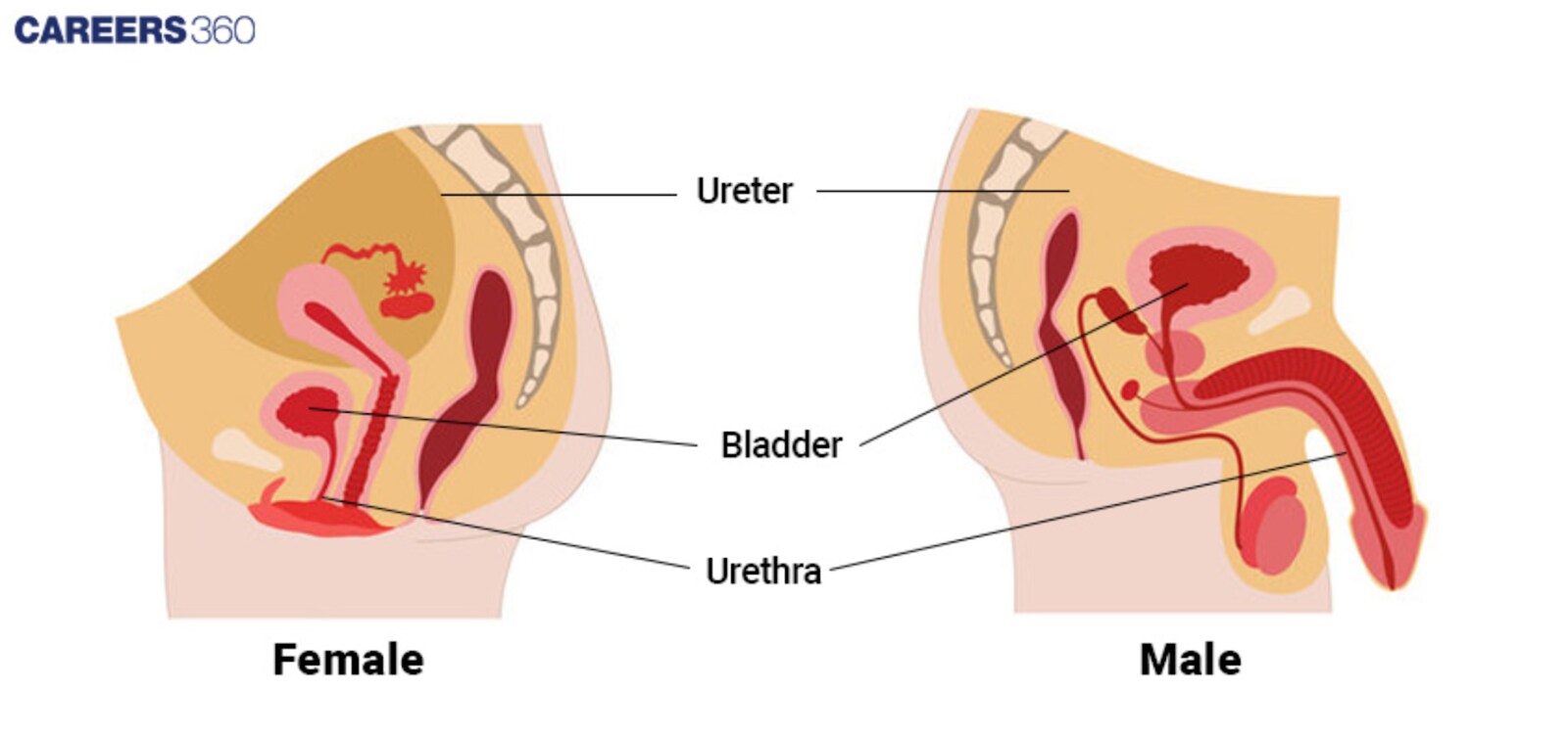
Urethra
The membranous urethra is a tubular organ that transmits urine from the urinary bladder to the outside of the body. It is different in terms of length and its anatomical course between males and females.
Description of the Male and Female Urethra
The male urethra is approximately 20 cm long.
In males, it performs the dual function of a passage for both urine and sperm.
It becomes partitioned into three portions:
Prostatic Urethra: It courses through the prostate gland.
Membranous Urethra: The shortest and narrowest portion, which runs through the urogenital diaphragm.
Spongy (Penile) Urethra: It courses along the entire length of the penis and opens at the external urethral orifice.
The internal urethral sphincter surrounds the male urethra at the neck of the bladder to prevent reflux of semen into the bladder at ejaculation.
Female Urethra:
The female urethra is about 4 cm long and functions only as a tube for the urinary passage.
It passes the length parallel to the wall of the vagina and opens into an orifice, that is the external urethral orifice lying in front of and separate from the vaginal orifice.
The length of the urethra in women is relatively short.
Thus, this increases the risk by which bacteria might reach the bladder and cause an infection, also known as a urinary tract infection or UTI.
Diagram of the Urethra
Function Of Urethra
The urethra performs the following functions:
Conduction of urine from the bladder to the outside of the body
The main role of the urethra is excretion, which refers to the process of expelling urine from the urinary bladder to the outside of the body, especially during the process of urinating. This is achieved by relaxing both the internal and external sphincters, which will readily allow the urine to flow through the urethra to the exterior of the body
Mechanisms of Urine Formation
Essentially, the overall processes concerned with urine formation within the kidneys can be summarized as glomerular filtration, tubular reabsorption, tubular secretion, and, lastly, excretion. Through these mechanisms, all waste products and excess substances are removed from the circulatory blood effectively while all essential nutritive factors and water are conserved.
Glomerular Filtration
Blood enters under high pressure within the glomerulus via the afferent arteriole, the vessel that perfuses the glomerulus.
It is the increased pressure in the glomerulus that forces out water, ions, and small molecules across the glomerular capillaries and into Bowman's capsule to form the glomerular filtrate.
This process clears waste products and excess substances from the blood, which then contribute to excretion. Thus urine formation begins
Factors Affecting Glomerular Filtration Rate (GFR)
Blood Pressure: An increase in blood pressure will increase GFR, while a decrease in it lowers it.
Afferent and Efferent Arteriolar Tone: alters these arterioles significantly alter GFR by giving rise to constriction or dilatation of these arterioles.
Plasma Protein Levels: The concentration of plasma proteins may alter the osmotic pressure responsible for drawing water and its contents back into the blood from the filtrate, thus affecting GFR.
Health of Glomerular Capillaries: Damage to or disease of the glomeruli will decrease filtration and lower GFR.
Tubular Reabsorption
Tubular reabsorption mainly occurs in the PCT, loop of Henle, DCT, and the collecting duct.
Proximal Convoluted Tubule: In the PCT, about 65-70% of the glomerular filtrate, water, ions, glucose, and amino acids get reabsorbed.
Henle's Loop: Here, water and sodium chloride are reabsorbed. It has a very vital role in concentrating urine.
PCT and Collecting Duct: The last adjustment in the amount of reabsorption of ions and water is according to body requirements due to aldosterone, antidiuretic hormone, and other hormones.
Tubular Secretion
Tubular secretion primarily takes place in the proximal convoluted tubule and the distal convoluted tubule.
Proximal Convoluted Tubule: There, it secretes waste products, such as hydrogen ions, ammonia, and certain drugs into the tubular fluid.
Distal Convoluted Tubule: Extra secretion of ions into Kramer, such as potassium and hydrogen, contributes to the maintenance of the body's acid-base balance.
Dialysis
Dialysis is a medical process used to simulate the functions of kidneys in patients whose kidneys become inadequate to filter by-products of metabolism from blood. It finds major indications in patients with ESRD, or end-stage renal dysfunction.
Also Read:
| Excretion | Urea Cycle |
| Glomerular Filtration Rate | Regulation of Kidney Function |
| Counter Current Mechanism | Hemodialysis |
Recommended Video on Parts of Human Excretory System
Frequently Asked Questions (FAQs)
The main constituents of the human excretory system are the kidneys, ureters, urinary bladder, and urethral passage. The kidneys filter blood to produce urine that afterwards passes into the ureters, enters the urinary bladder, and in due time gets excreted out of the body via the urethra.
Blood is filtered by the kidneys using glomerular filtration. Blood enters the glomerulus, a cluster of capillaries within the renal corpuscle. The increased blood pressure within the glomerulus forces the water, ions, and small solutes through its capillary walls into Bowman's capsule, thus making the glomerular filtrate. The filtered solution undergoes both reabsorption and secretion through the renal tubules to become the urine.
The ureters are muscular tubes that carry urine from the kidneys to the urinary bladder. In this way, the peristaltic contractions of the walls make the ureters propel the urine downwards and hence make its way very smooth and also prevent backflow.
Common pathologies to the excretory system include renal calculi, UTIs, CKD, AKI, and disorders of the bladder like interstitial cystitis and incontinence. These pathologies can further result in damage to its role of filtering wastes and regulating fluid balance.
A good excretory system is maintained by keeping the body well-hydrated, consuming a healthy diet with low levels of sodium and processed foods, working out regularly, avoiding excessive alcohol and caffeine consumption, and not smoking at all. This permits regular checkups with health providers to monitor kidney function, allowing for problems to be identified early on.
Also Read
28 Nov'24 08:16 PM
28 Nov'24 06:24 PM
28 Nov'24 12:06 PM
27 Nov'24 04:33 PM
27 Nov'24 02:40 PM
27 Nov'24 12:24 PM
Articles

Student Community: Where Questions Find Answers