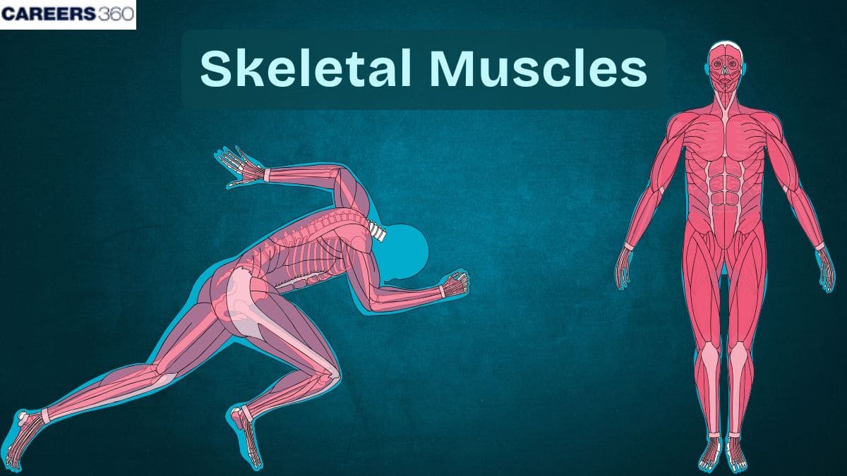
Skeletal Muscles: What Is It, Function, Location, Anatomy
Skeletal muscles are striated, voluntary tissues responsible for producing body movements through contraction, posture maintenance, heat production, and organ protection. Their highly organized structure—myofibrils, actin–myosin filaments, sarcomeres, connective tissues, and rich innervation—enables efficient and controlled contraction. This NEET-focused guide covers skeletal muscle definition, structure, types of fibres, properties, functions, disorders, diagrams, FAQs & MCQs.
This Story also Contains
- Definition of Skeletal Muscles
- Structure of Skeletal Muscles
- Function of Skeletal Muscles
- Types of Skeletal Muscle Fibers
- Comparison of Skeletal, Smooth & Cardiac Muscles
- Disorders Related to Skeletal Muscles
- Skeletal Muscles NEET MCQs (With Answers & Explanations)
- Recommended Video on "Skeletal Muscle"

Definition of Skeletal Muscles
Skeletal muscles are striated, voluntary tissues of muscles attached to bones by tendons and responsible for initiating or controlling body movements. These muscles have long, cylindrical fibres and striations due to actin and myosin filaments realizing activities right from locomotion to the maintenance of posture or heat production.
Apart from the skeletal, there are other major divisions of muscles: cardiac muscle, which is involuntary and found in the heart and smooth muscle, which is involuntary, located within the walls of internal organs. All three types of muscles combine to perform very vital functions necessary for the overall operations of the body.
Structure of Skeletal Muscles
The structure of the skeletal muscles is defined as:
Muscle Fibre (Cell)
Each fibre of the external, striated skeletal muscle is, in reality, a rather long, cylindrical cell having several nuclei at its periphery. Such fibres are truly multi-nucleated and are covered by a plasma membrane known as sarcolemma. The latter forms a wrapper for the cytoplasm called the sarcoplasm.
Myofibrils & Myofilaments
These muscle fibres are covered under myofibrils, which in turn are composed of filaments called myofilaments. Myofilaments consist of actin and myosin, which interact to bring about muscle contraction. The pattern of these filaments inside the muscle gives it a striated appearance.
Sarcomere – Functional Unit
It is the functional unit of muscle contraction.It is technically the segment between two Z adjacent discs. Where overlapping in actin and myosin filaments occurs, which contracts and relaxes by extending and shortening to make a muscle move.
Connective Tissue Layers
The components of the connective tissue are:
Endomysium: This is the thin connective tissue surrounding every individual fibre. These also provide structural support and include capillaries and nerves collecting and supplying the muscle fibres respectively.
Perimysium: This is the connective tissue sheath enclosing bundles of muscle fibres the fascicles. It also gives some mechanical support, and it includes larger blood vessels and nerves than the endomysium does.
Epimysium: It refers to the external connective tissue to the whole muscle. It is continuous with the tendon, and thus forms a protective sheath for the muscle as such and renders some structural integrity.
Blood Supply & Innervation
The blood supply to the skeletal muscles is very rich, with an extensive network of capillaries furnishing the oxygen and nutrient requirements for proper muscle functioning.
Muscle fibres are also innervated by nerves, especially motor neurons, that aid in the voluntary control and coordination of each muscle contraction.
Function of Skeletal Muscles
The skeletal muscle performs the following functions:
Movement
Skeletal muscles of the body originate and control most of their voluntary movements.
This is achieved through the simple act of muscle contraction, thus causing the bone to be pulled, which acts as leverage leading to movement around the joints.
These can be simple actions like walking or picking up something too highly intricate from the playing of musical instruments to sports.
Posture & Support
The other major functions of the skeletal muscles, in conjunction with the provision for movement, include posture and support.
These muscles have a continuous action in counteracting the force of gravity in holding up the body in an upright position and balance.
Heat Production
Skeletal muscle contractions produce heat through metabolic by-products of muscle contractions and play a role in the maintenance of core body temperature.
When exercising or exposed to low temperatures, thermogenesis increases the metabolic rate to maintain the inner temperature through enhanced production of heat as a result of muscle contractions.
Protection of Organs
The skeletal muscles also protect the internal organs through protective mechanisms and additional structural support.
The abdominal wall and thoracic cavity muscles safeguard vital organs like the intestines, heart, and lungs from shock and other forms of physical trauma.
This protective function assumes paramount importance in the preservation of the integrity and safety of these internal systems.
Types of Skeletal Muscle Fibers
The different types of skeletal muscle fibres are:
Red (Slow-Twitch, Type I)
The red muscle fibres or Type I or slow-twitch fibres, are rich in myoglobin, generally giving them a red colour.
They have a high density of mitochondria and capillaries.
Thus, having a high amount of mitochondria inside the cell, they can efficiently produce energy in the form of aerobic metabolism.
For that reason, the fibres are made for endurance as well as prolonged activities, such as running a long distance or simply maintaining posture by continued contraction without developing fatigue.
White (Fast-Twitch, Type II)
White muscle fibres contain few myoglobins and a few mitochondria, hence their pale colour.
They depend more on anaerobic metabolism for the production of energy, so they can produce rapid and powerful contractions.
Hence, they would do better under brief, intense activities like sprinting or weightlifting.
They are capable of applying great forces but tire more quickly than red muscles.
Comparison of Skeletal, Smooth & Cardiac Muscles
The difference between skeletal, smooth and cardiac muscle is discussed in the table below:
Feature | Skeletal | Smooth | Cardiac |
Control | Voluntary | Involuntary | Involuntary |
Striations | Present | Absent | Present |
Nuclei | Multi | Single | Single |
Location | Attached to bones | Hollow organs | Heart |
Contraction | Fast | Slow, sustained | Rhythmic |
Disorders Related to Skeletal Muscles
Several disorders and diseases can compromise muscle and thus function and quality of life.
Muscular Dystrophy
Genetic mutations that alter muscle proteins.
Treatment is symptomatic and aims to retard the progression.
Duchenne muscular dystrophy: Progressive muscle weakness
Becker muscular dystrophy: Similar but milder
Myasthenia Gravis
Muscle weakness, fatigue
Autoimmune disorder at the neuromuscular junction.
Medications aimed at improving nerve-muscle communication.
Immunosuppressive therapies.
Muscle Cramps and Strains
It is caused by dehydration, overuse, and electrolyte imbalance.
This can be prevented by regular stretching and proper hydration.
Rest, ice application, compression, elevation (RICE)
Gentle stretch and rehydrate
Skeletal Muscles NEET MCQs (With Answers & Explanations)
Important questions asked in NEET from this topic are:
Structure & function of skeletal muscles
Skeletal vs Smooth vs Cardiac muscles
Disorders related to muscles
Practice Questions for NEET
Q1. The dark band present on myofibril is
Isotropic band
Anisotropic band
Hensen's zone
M-line
Correct answer: 2) Anisotropic band
Explanation:
One of the darker bands in a muscle fiber, known as the A-band, consists primarily of myosin filaments. In this region, one can observe the arrangement of myosin proteins since they are placed in rod-like structures that run parallel to each other and along the longitudinal axis of the myofibrils. This A-band is very important in the contraction process of muscles because it makes contact with actin filaments during the sliding filament mechanism which may contribute to increased muscle strength and functionality.
Hence, the correct answer is option 2) Anisotropic band.
Q2. The plasma membrane of the muscle fiber is called
Sarcoplasma
Sarcolemma
Sarcoplasmic Reticulum
Syncytial
Correct answer: 2) Sarcolemma
The plasma membrane in a muscle fibre, or muscle cell, is termed the sarcolemma. This crucial biological barrier performs dual functions: preserving the muscle fibre's integrity and facilitating the conduction of electrical signals. The sarcolemma's primary role lies in the transmission of action potentials, which are essential electrical impulses that initiate muscle contraction. It features specialized components, like t-tubules, which are responsible for efficiently conducting these impulses throughout the muscle fibre. T-tubules are transverse tubules that extend from the surface to the interior, allowing the action potential to reach the necessary depth within the muscle fibre. The sarcolemma's integrity is vital for the correct operation of muscle contraction mechanisms and for maintaining communication between the cell's exterior and its internal components.
Hence, the correct answer is option 2) Sarcolemma.
Q3. The muscle fibers that contract slowly are
Red muscle fibres
White muscle fibres
Both a and b
None of these
Correct answer: 1) Red muscle fibres
Explanation:
Red muscle fibers, also known as slow-twitch fibers, contract slowly but are highly resistant to fatigue. They are rich in myoglobin, mitochondria, and blood supply, which enable sustained aerobic energy production. These fibers are well-suited for endurance activities like walking, running long distances, or maintaining posture, as they rely on oxidative metabolism for energy.
Hence, the correct answer is option 1) Red muscle fibers.
Also Read:
Recommended Video on "Skeletal Muscle"
Frequently Asked Questions (FAQs)
The contraction of a muscle originates from myosin filaments that pull on actin filaments, causing them to slide towards the centre of the sarcomere, effectively shortening the muscle. It necessitates ATP and an initiating signal from the nervous system.
Slow-twitch fibres are endurance-oriented; therefore, high in myoglobin, and thus resistant to fatigue; fast-twitch fibre types are stranded or fast with low myoglobin and quick to fatigue.
Skeletal muscle disorders include dystrophies of muscle—Duchenne muscular dystrophy, for example—myasthenia gravis, and strains or tears of muscles.
Exercise strengthens and increases the durability of muscle, while a well-balanced diet supplies its restoration and development with the appropriate kinds and amounts of proteins and other nutrients.
Skeletal muscles are striated, involuntary muscles inelastically attached to bones. Skeletal muscles mediate movement, maintenance of posture, generation of heat, and protection of internal organs.