

Vertebral Column: Function, Anatomy, Structure, Topic
The vertebral column, or backbone, forms the central supporting axis of the human body and replaces the embryonic notochord. Composed of 33 vertebrae (26 in adults due to fusion), it protects the spinal cord and allows flexibility, posture, and movement. This guide explains the anatomy, regions, structure of vertebrae, functions, common disorders, diagrams, FAQs, and NEET MCQs.
This Story also Contains
- What Is the Vertebral Column?
- What Are Vertebrae?
- Anatomy Of The Vertebral Column
- Structure Of Vertebrae
- Functions Of The Vertebral Column
- Common Disorders Of Vertebral Column
- Vertebral Column NEET MCQs (With Answers & Explanations)
- Recommended Video for Vertebral Column
What Is the Vertebral Column?
The vertebral column is also known as the spine or backbone of the human body. This column made up of 26 small vertebrae provides support, protection, and flexibility for movement in the adult human body.
What Are Vertebrae?
An adult spine has 26 vertebrae, while the total amount of vertebrae is 33. This is so because the sacral and the coccygeal vertebrae fuse to form one. The vertebral column replaces the notochord from the embryonic stage and makes up the primary structure of the trunk attaching at the base of the skull. Each vertebra has a hollow forming the neural canal through which the spinal cord passes thus protecting it.
Anatomy Of The Vertebral Column
The human spine is divided into five sections, all of which are different in character and functions. These include the following:
Cervical Spine (7 vertebrae)
The first vertebra, C1 or Atlas, articulates with the skull. This occurs with the 'yes' movement of the head.
The second vertebra, C2 or Axis, with C1, provides the 'no' movement of the head.
It supports the weight of the head.
Thoracic Spine (12 Vertebrae)
Still relatively less mobility, holds up the rib cage, which protects the lungs and heart.
The thoracic vertebrae articulate with the 12 pairs of ribs.
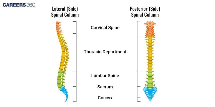
Lumbar Spine (5 vertebrae)
Holds the weight of the body.
These are large, hence one can carry a heavyweight.
Sacral Spine (5 Fused Vertebrae)
Fused five sacral vertebrae
Joins the vertebral column to the hip bones and thus forms the pelvic girdle
Coccygeal Spine ( 4 Fused Vertebrae)
Four coccygeal vertebrae fuse to form the tailbone.
These supports sitting posture
Structure Of Vertebrae
The structure differs from one region of the vertebra to another. The general or typical vertebra is made up of the following parts:
Vertebral body
Vertebral arch
Vertebral foramen, forming the spinal canal
Processes: spinous, transverse, and articular
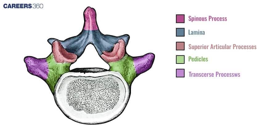
Each of the vertebrae is held together by several ligaments, thus promoting movement and stability. The hollow cavity that is formed by the central portion of every vertebra encloses the spinal cord and thus protects it.
Functions Of The Vertebral Column
The functions of the vertebral column include:
Support
The vertebral column provides for the structural support of the body, thus supporting the head and trunk.
Protection
Protection of the spinal cord and nerves within the vertebral canal.
Movement
Allows flexibility, hence allowing bending and twisting within the range of motion.
Attachment
Muscle and ligament attachments in the human body.
Common Disorders Of Vertebral Column
Damage to any of the vertebrae or their parts can lead to a lot of complications and limitation of movement. The common disorders are
Scoliosis
Abnormal sideways curvature of the spine
Appears mostly during childhood or adolescence
Unevenness in shoulders, waist, and hips
Serious cases can affect lung and heart
Treatment includes observation, bracing, surgery
Herniated Disk
Also called slipped disk or ruptured disk
When the inner gel of a spinal disk protrudes through a tear in the outer layer
Nerve compression causing pain, numbness, or weakness
Common in the lower back but most frequent in the neck
Treatment includes physical therapy, medication, surgical intervention
Osteoporosis
Characterised by brittle, porous, and weakened bones
Common in elderly people, especially postmenopausal women
Increase risk of fractures from minor falls/ simple actions
Diet rich in calcium and vitamin D, regular exercises, medications for stronger bones
Spinal Stenosis
Narrowing of spaces within the spine that puts pressure on the spinal cord and nerves
Majorly present in the lower back and neck
Symptoms are pain, numbness, muscle weakness, problems with bladder or bowel control
Usually caused by age-related changes such as thickened ligaments, bone spurs, or herniated discs
Treated with physical therapy, medication, or even surgery
Vertebral Column NEET MCQs (With Answers & Explanations)
Important questions asked in NEET from this topic are:
Anatomy of the vertebral column
Functions of the vertebral column
Practice Questions for NEET
Q1. In humans, the coccyx, also known as the tailbone, is formed by the fusion of multiple vertebrae. Among the following options, determine the accurate number of vertebrae involved in this fusion:
Three vertebrae
Four vertebrae
Five vertebrae
Six vertebrae
Correct answer: 2) Four vertebrae
Explanation:
The coccyx in humans is formed by the fusion of four vertebrae. It is the remnant of a tail that our ancestors had. The fusion of these four small vertebrae creates the triangular-shaped coccyx, located at the bottom of the vertebral column.
Hence, the correct answer is option 2) Four vertebrae
Q2. The cervical vertebra called the axis provides the head with sideways rotation. This can be because
It is articulated to skull through occipital condyles
It is fused with 1st vertebra atlas
It is joined through elastic pads of fibrocartilage with other vertebrae, which provide mobility
It contains odontoid process that fits into the odontoid canal of atlas
Correct answer: 4) It contains odontoid process that fits into the odontoid canal of atlas
Explanation:
The cervical vertebra called the axis (C2) allows the head to rotate sideways due to the presence of a unique structure called the dens or odontoid process. This peg-like projection extends upward and fits into the vertebra above it, the atlas (C1), forming the atlantoaxial joint.
Hence, the correct answer is option 4) It contains an odontoid process that fits into the odontoid canal of the atlas.
Q3. Which knob-like process on the second cervical vertebra in human beings is responsible for rotational movements of the head?
Prezygapophysis
Postzygapophysis
Olecranon process
Odontoid process
Correct answer: 4) Odontoid process
Explanation:
The odontoid process is located on the second cervical vertebra and acts as a knob-like projection that enables rotational movements of the head. It serves as a pivotal point for the rotation between the first cervical vertebra (atlas) and the head. Essentially, the odontoid process allows for the turning and twisting motions of the head.
Hence, the correct answer is option 4) Odontoid process.
Also Read:
Recommended Video for Vertebral Column
Frequently Asked Questions (FAQs)
Human beings have a total of 33 vertebrae during development, but in adults, the number is 26 because some vertebrae fuse. icans have a total of 33 vertebrae during development, but in adults, there are 26 because some vertebrae fuse.
The four main functions include protection of the spinal cord, support of the head, anchorage of the ribs and muscles, and locomotion with movement.
The Greeks called the first one Atlas (C1) and the second Axis (C2).
The bones of the spine, which are 26 in number, are called vertebrae. They are cervical, thoracic, lumbar, sacral, and coccygeal vertebrae.
Five divisions include the cervical, thoracic, lumbar, sacral, and coccygeal.
