

Continuous X-ray
Continuous X-rays, a form of electromagnetic radiation, play a pivotal role in various fields of science and medicine. Unlike their more commonly known counterparts, the discrete X-rays, continuous X-rays are generated by the rapid deceleration of high-energy electrons, resulting in a spectrum of X-ray wavelengths. This phenomenon is fundamental to medical imaging techniques, particularly in diagnostic radiology, where it enables detailed internal views of the human body. For instance, continuous X-rays are used in CT scans to provide cross-sectional images of bones, organs, and tissues, aiding in the diagnosis of conditions ranging from fractures to tumours. Beyond medicine, continuous X-rays are also employed in security scanners at airports, ensuring the safety of travellers by detecting concealed objects in luggage. Their application extends to industrial non-destructive testing, where they help in examining the integrity of materials and structures without causing damage. Through these real-life examples, the importance of continuous X-rays in enhancing our understanding and safety in everyday life becomes evident. In this article, we will discuss the concept of continuous X-rays and provide examples for better understanding.
This Story also Contains
- Solved Examples Based on Continuous X-ray
- Example 1: Electrons with an energy of 80 keV are incident on the tungsten target of an X-ray tube. K-shell electrons of tungsten have -72.5 keV energy. X-rays emitted by the tube contain only,
- Summary
Continuous X-ray
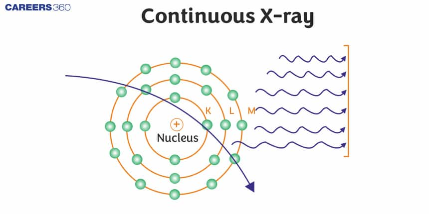
As we know about the phenomenon of visible light, similarly continuous X-ray spectra also contain photons ranging through a lot of wavelengths. From the previous concept, we know that the production of X-rays happens when the target which is made up of an element with a high atomic number is hit by electrons traveling at a high velocity. So out of the total energy, most of the energy applied is wasted by being converted into heat energy in the target material’s system. X-rays that have continuously unstable wavelengths are produced due to the loss of energy that the few electrons that were moving fast enough (and penetrated to the interior sections of the atoms of the material being targeted) suffer. Since the attractive pulling forces applied by the nucleus of the target element cause a deceleration of these fast-moving electrons, this continuously decreases the energy of the electron. Due to this, varying frequency of X-rays is emitted continuously due to the retardation of the speed of electrons. The X–rays consist of a continuous range of frequencies up to a maximum frequency $\nu_{\max }$ or minimum wavelength $\lambda_{\text {min }}$. This is called continuous X–rays. The minimum wavelength depends on the anode voltage. If V is the potential difference between the anode and the cathode, then -
$e V=h \nu_{\max }=\frac{h c}{\lambda_{\min }}$
To produce the continuous X-ray in the Coolidge tube, an electron is projected toward the anode with an accelerating voltage V. So, the kinetic energy of the projectile electron will be eV. As shown in the figure, it experiences strong electric force toward the nucleus of the atom and due to this strong attraction the velocity of this electron, when it emerges from the atom, will be highly reduced and negligible compared with the initial speed of the projectile electron.

According to the law of conservation of energy, the energy of these electromagnetic radiations will be equal to the decrease in the kinetic energy of the projectile electron.
$\begin{aligned} & e V=\frac{1}{2} m v^2 \\ & v=\sqrt{\frac{2 e V}{m}}\end{aligned}$
However, the velocity of the incoming electron will be less than that of the projectile electron. This difference in kinetic energy will cause the production of X-rays.
Recommended Topic Video
Solved Examples Based on Continuous X-ray
Example 1: Electrons with an energy of 80 keV are incident on the tungsten target of an X-ray tube. K-shell electrons of tungsten have -72.5 keV energy. X-rays emitted by the tube contain only,
1) A continuous X-ray spectrum with a minimum wavelength of ≈ 0.155$\AA$
2) A continuous X-ray spectrum (Bremsstrahlung) with all wavelengths
3) The characteristic X-ray spectrum of tungsten
4) A continuous X-ray spectrum (bremsstrahlung) with a minimum wavelength of $\approx 0.0155 \mathrm{~nm}$ and the characteristic X-ray spectrum of tungsten
Solution:
The minimum wavelength of continuous spectrum
$\begin{aligned} \lambda & =\mathrm{hc} / \mathrm{E} \\ \lambda & =\frac{6.6 \times 10^{-34} \times 3 \times 10^8}{80 \times 10^3 \times 1.6 \times 10^{-19}}\end{aligned}$
Apart from continuous X-rays, as the energy of the incident electrons is greater than the magnitude of the energy of the K-shell electrons, the target atoms will have vacancies in the K shell (K-shell electrons will be knocked out). This will cause the emission of the entire characteristic spectrum of tungsten.
Hence, the answer is the option (4).
Example 2: An X-ray tube is operated at 1.24 million volts. The shortest wavelength of the produced photon will be :
1) 10 -2 nm
2) 10 - 1 nm
3) 10 -4 nm
4) 10 -3 nm
Solution:
The minimum wavelength of the photon will correspond to the maximum energy due to accelerating by V volts in the tube.
$
\begin{aligned}
& \mathrm{eV}=\frac{h c}{\lambda_{\min }} \\
& \lambda_{\min }=\frac{\mathrm{hc}}{\mathrm{eV}} \\
& \lambda_{\min }=\frac{1240 \mathrm{~nm}-\mathrm{eV}}{1.24 \times 10^6} \\
& \lambda_{\min }=10^{-3} \mathrm{~nm}
\end{aligned}
$
Hence, the answer is the option (4).
Example 3: An electron having de-Broglie wavelength $\lambda$ is incident on a target in an X-ray tube. The cut-off wavelength of the emitted X-ray is:
1) 0
2) $\frac{2 \mathrm{~m}^2 \mathrm{c}^2 \lambda^2}{\mathrm{~h}^2}$
3) $\frac{2 m c \lambda^2}{h}$
4) $\frac{h c}{m c}$
Solution:
For cut-off length, the total kE of moving electron is dissipated to radiate x-ray
$\begin{aligned} & \lambda_{\text {electron }}=\frac{h}{m v}= \frac{h}{\sqrt{2 m k E}} \\ & \lambda^2=\frac{h^2}{2 m(K E)}\end{aligned}$
$\lambda_{\text {cutoff }}=\frac{h c}{(k E)}=\frac{h c}{\frac{h^2}{2 m \lambda^2}}=\frac{2 m c \lambda^2}{h}$
Hence, the answer is the option (3).
Example 4: The $\kappa_\alpha$ X-ray emission line of tungsten occurs at $\lambda=0.021 \mathrm{~nm}$. The energy difference ( in KeV ) between K and L levels in this atom is:
1) 59
2) 49
3) 3.9
4) 69
Solution:
Different types of characteristics of X-ray
$\begin{aligned} & K_\alpha(L \rightarrow K) \text { Transition } \\ & K_\beta(M \rightarrow K) \text { Transition } \\ & K_\gamma(N \rightarrow K) \text { Transition } \\ & L_\alpha(M \rightarrow L) \text { Transition } \\ & L_\beta(N \rightarrow L) \text { Transition } \\ & M_\alpha(N \rightarrow M) \text { Transition } \\ & E_K-E_L=\Delta E_{K \alpha}=\frac{h c}{\lambda}=\frac{6.62 \times 10^{-34} \times 3 \times 10^8}{0.021 * 10^{-9}} \\ & \Delta E=59 \mathrm{KeV}\end{aligned}$
Hence, the answer is the option (1).
Summary
Continuous X-rays, generated by the rapid deceleration of high-energy electrons, form a crucial part of medical imaging, security scanning, and industrial testing. They produce a spectrum of X-ray wavelengths essential for detailed internal imaging in CT scans and other applications. Through examples and problem-solving, the concepts of continuous X-ray production and their real-life applications are better understood, highlighting their significance in technology and safety measures.
