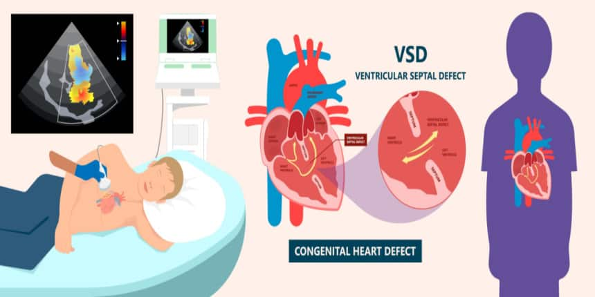
VSD FULL FORM
What is the full form of VSD?
VSD stands for Ventricular Septal Defect. Commonly people call VSD “a hole in the heart”. It is a type of defect in the ventricular septum or the wall which separates chambers, and ventricles from each other. VSD is usually present at birth but with no visible symptoms. It is commonly found in newborns and infants. Rarely do older children and adults have VSDs. VSD is one of the most common cardiac abnormalities known to humans. Of all the newborns, 30-60% are born with this VSD in the world. It is one of the most common congenital heart defects found in humans.
- What is the full form of VSD?
- What is Ventricular Septal Defect(VSD)?
- Causes of Ventricular Septal Defect(VSD)?
- Symptoms of Ventricular Septal Defect(VSD)?
- Diagnosis of Ventricular Septal Defect(VSD)?
- Treatment of Ventricular Septal Defect(VSD)?

What is Ventricular Septal Defect(VSD)?
Ventricular Septal Defect changes blood circulation flow in the heart which ultimately results in the change of blood flow through the lungs as well. A VSD disrupts the piping ability of the heart because of the leak or the hole in the wall. This leak can cause extra blood in the lungs which can further increase the problem for the patient.
A VSD changes the flow of blood circulation in the heart and lungs of the patient. It makes oxygen-rich blood from the heart to the lungs instead of pumping out of the body. Hence, oxygen-rich and poor blood get mixed together. This increases blood pressure in the lungs due to which the heart has to pump out blood with more force. Thus, this creates congestion in the blood arteries of the lung.
Causes of Ventricular Septal Defect(VSD)?
Ventricular Septal Defects (VSD) causes are not yet known to the world. But genetics and environmental issues do play a significant role in causing the defect. This defect is often associated with other congenital defects.
Many times VCD is caused during embryonic stages, particularly in the fifth week of the gestation period. As the fetus develops, the heart doesn’t have separate ventricles. Later a wall which is also known as the septum wall is developed which separates the right and left ventricles. Whenever the wall is not completely formed, a hole develops i.e. the ventricular septal defect. The number of holes and sizes can vary from person to person.
Whenever VSD arises due to genetic disorders it can be extra or missing chromosomes.
A VSD can also arise after a few days of heart attack due to the tearing of the septal wall. This only happens before white blood cells start functioning in the dead tissues of the heart to form the scar tissues.
Symptoms of Ventricular Septal Defect(VSD)?
Commonly Ventricular Septal Defects (VSD) are found in infants, but there are a few cases where it was diagnosed in older children and adults as well.
Symptoms can vary depending on the number of holes and sizes of the holes in an individual. Many times people with VSD may not notice any symptoms but doctors can identify them by the murmur of the heart.
Most of the common symptoms in the infants are following:
Breathing issues like shortness of breath fast breathing or struggle to breathe
During feeding, infants may be sweating
Fatigue especially during feeding
Frequent respiratory problems
Slow weight gain
Most of the common symptoms in older children and adults are the following:
During exercising they may experience tiredness or shortness of breath
Heart inflammation due to infections
Pale or bluish skin and lips
The trouble with weight gain
Lack of appetite or a very poor one
Diagnosis of Ventricular Septal Defect(VSD)?
Ventricular Septal Defect(VSD) is generally diagnosed by doctors when they hear a heart murmur using the stethoscope. Doctors can even estimate the size of the VSD from the sound of a murmur.
Many times during physical exams VSD can miss due to being very small. But there are many imaging tests for detecting the hole and its size.
Echocardiogram: It is a painless and fast test to check VSD defects in the heart. This form an image with the help of ultra-high frequency sound waves. This image shows the inside and outside of the heart which helps in locating the VSD and its size.
Electrocardiogram: This is also commonly known as ECG or EKG. It has multiple sensors to be attached to the chest to detect VSD. The heart activity is recorded in print or digital form as a wave. Any disturbance in this wave is due to the defect. But many times ECG fails to detect a VSD if the heart hasn’t begun to change its shape or the size of the VSD is too small for electrical activity.
Chest or heart X-ray: This test is used when VSD is too large, as the shape of a heart is changed or has already begun to change. In some cases, a substance is used to show unusual blood circulation on the X-ray. This substance is injected into the blood very carefully by healthcare providers.
Computed Tomography Scan: This is commonly known as CT Scan. It is a test where a 3D or three-dimensional image of the heart is generated. In this, also a substance is injected into the blood to make it visible on the scan which helps in determining the VSD.
Cardiac catheterization: In this, a small device i.e. catheter is inserted into the blood vessel to look into the inside of the heart. This is generally advised for patients with lung problems. The catheter is inserted from the upper thigh and passes up to the heart to locate the hole and its size.
Treatment of Ventricular Septal Defect(VSD)?
Treatment of the VSD depends on the age of the patient, and size of the hole, and the location of the hole on the wall between the ventricles. Most of the Ventricular Septal Defects (VSD) are too small and close on their own by the age of 6 in small children which causes hardly any health problems. So healthcare advises in these cases to avoid any surgery and see for time being that the defect closes itself with time.
But when VSDs are big or diagnosed later age, doctors advised the following treatments:
Surgery: It is the most surgical method to treat VSDs. In this, a patch is used to close the hole. The patch used in this can be a synthetical material piece or a graft of the patient’s tissue. After six months of the surgery, the patch or the tissue will become a part of the wall of the heart between the ventricles.
Transcatheter procedure: In this method, a catheter is inserted from a major artery to plug the hole by using an occluder. The device ultimately will become a part of the wall between ventricles as the tissue of the heart will grow around the occluding device.
Rare Complications of Ventricular Septal Defect(VSD)?
Ventricular Septal Defect(VSD)
Arotic insufficiency:
Damage to the heart causing irregular heart rhythm
Lack of growth and development
Heart Failure
Heart Bacteria Infections
High BP in the lungs can cause the failure of the heart’s right side
Premature birth of the baby
Down syndrome and genetic conditions
Atrial septal defects
Coarctation of the aorta
Double outlet syndrome
Patent ductus arterious
Tetralogy
Eisenmenger syndrome
Endocarditis
Prevention of Ventricular Septal Defect(VSD)?
Ventricular Septal Defects (VSD) are generally present from birth but rarely they arise after a cardiac or heart attack.
There is no certain prevention for VSD but taking the following precautions during pregnancy can definitely lower the risk of VSD in the baby.
Avoiding alcohol consumption
Using antiseizure medicines can increase the risk
Getting early parental care
Taking folic acids with multivitamins
Don’t consume illegal drugs or smoke
Take all vaccinations recommended by the doctors
Keeping diabetes in check
Frequently Asked Questions (FAQs)
The full form of VSD is Ventricular Septal Defects.
The exact cause of VSD is still not known, but few theories are there.
There is no prevention for VSD.
Doctors are known as cardiologists.