Difference Between Arteries and Veins
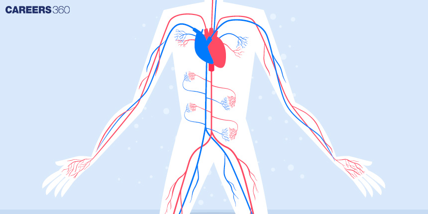
Arteries and veins are essential components of the circulatory system. Arteries carry oxygen-rich blood away from the heart to various body parts, while veins return oxygen-depleted blood to the heart. These blood vessels have distinct structures and functions, ensuring efficient blood flow throughout the body. In this article, Arteries and Veins, Structure of Arteries And Veins, Types of Arteries and Veins, Characteristics of Arteries, Characteristics of Veins, and Key Differences between Arteries and Veins are discussed. Arteries and Veins are a topic of the chapter Body Fluids and Circulation in Biology.
NEET 2025: Mock Test Series | Syllabus | High Scoring Topics | PYQs
NEET Important PYQ's Subject wise: Physics | Chemistry | Biology
New: Meet Careers360 B.Tech/NEET Experts in your City | Book your Seat now
- What are Arteries and Veins?
- Structure of Arteries And Veins
- Types of Arteries and Veins
- Characteristics of Arteries
- Characteristics of Veins
- Key Differences between Arteries and Veins
What are Arteries and Veins?
Blood vessels are part of the Circulatory system which includes arteries, and veins these are blood vessels that transport blood all over the body and capillaries. Arteries are blood vessels that transport well-oxygenated blood from the heart to different parts of the body whereas veins are the vessels that transport poorly oxygenated blood back to the heart. Both of these types of vessels are essential for adequate blood flow, the delivery of oxygen and nutrients to the tissues and the removal of waste products from these tissues. An overview of arteries and veins is therefore important in understanding the concept of circulatory systems and the significance of vascular health.
Also Read-
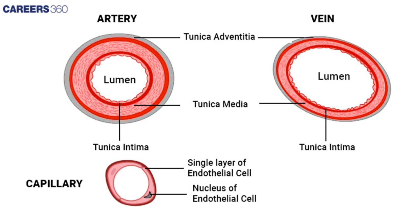
Structure of Arteries And Veins
The structure of arteries and veins is described below:
Arteries
The structure of arteries is described below:
Thick, muscular walls
Arteries have thick walls made up of three tunics: tunica intima, which is the inner layer of flat endothelial cells; tunica media which could be a muscular and elastic layer as found in large arteries; and tunica externa the outermost connective layer. Large muscular walls guard and control high blood pressure resulting from the blood pumped from the heart.
Elastic fibres
Some important elements of these fibres are required to give proper flexibility to arteries for each contraction and relaxation during the heartbeat to manage proper blood flow.
Narrow lumen
The arteries hence have a relatively small inner diameter known as the lumen hence the ability to withstand high pressure to enable blood to flow nicely.
Presence of smooth muscle cells
These cells that are located in the tunica media control the contraction and dilation of arteries to control blood pressure and the rates of flow of blood in the body.
Veins
The structure of veins is described below:
Thin walls
Compared to the arteries, veins are vessels of smaller diameter, they do not have much musculature and elastic tissue because they pump the blood under lower pressure.
Less elastic tissue
The elasticity is less in the veins this is because veins return blood at low pressure to the heart.
Wider lumen
Veins are blood vessels with slightly wider lumen to contain a significantly larger amount of blood that will be pumped back to the heart.
Valves to prevent backflow
Blood vessels like the veins that are found in the legs part have valves which prevent the backflow of blood and the blood will only flow in one direction to the heart.
Types of Arteries and Veins
The types of arteries and veins are described below:
Types of Arteries
The types of arteries are:
Elastic Arteries
Description: These are the largest arteries nearest to the heart and they have thick walls with greater proportionality of elastic fibres. This elasticity enables them to receive blood when there is an increase in pressure as a result of pumping from the heart and ensures normal flow of blood.
Examples: Pulmonary artery and the aorta.
Muscular Arteries
Description: These arteries contain a proportionately larger quantity of smooth muscles rather than elastic fibres, through which they can regulate the blood flow by changing the diameter of the blood vessels. They transport blood to other organs of the body.
Examples: The radial artery and the femoral artery.
Arterioles
Description: They are tiny blood vessels that regulate the quantity of blood to be delivered to an area of the body. It includes a rather feeble muscular layer compared to its measures and they have chances to control blood flow discreetly and alter the pressure.
Function: They act as pressure maintaining and directing vessels whereby they maintain blood pressure and then direct the circulations into capillary beds.
Types of Veins
The types of veins are:
Superficial veins
Description: These veins are near the dermis and therefore they are easily seen on the skin. They allow blood and fluid to be drawn out of the skin and the first layers of the tissue.
Examples: These are the greater saphenous vein and the small saphenous vein.
Deep veins
Description: Located more towards the centre and frequently in association with arteries deep veins transport the blood from internal organs and muscles and back to the heart. They are more crucial for blood flow and pressure maintenance.
Examples: The femoral vein provided the major contribution to the popliteal vein while the brachial vein contributed to the antebrachial vein.
Venules
Description: These are the smallest blood veins and take blood deposits from the tiny capillaries and these accumulate in other bigger veins. It is thin-walled and is engaged in the process of collecting blood from the capillary beds.
Function: Capillaries are small blood vessels which allow blood to flow from arterioles to venues and thus play a very important role in circulation by channelling blood back to the veins.
Characteristics of Arteries
1. Situated deep into a muscle
2. Have extremely thick walls
3. Transfer blood between the organs and the heart.
4. Deliver oxygen-rich blood (except for the pulmonary artery)
5. Internally has a thick layer of muscular tissue
6. Lack valves (except for the pulmonary artery)
Characteristics of Veins
1. Are situated closer to your body's surface.
2. Possess thin walls
3. Direct blood flow to your heart
4. Carry anaemic blood
5. Contain a thin coating of muscular tissue
6. Have valves to maintain blood flow
Key Differences between Arteries and Veins
It is one of the important difference and comparison articles in biology. The differences are listed below-
Aspect | Arteries | Veins |
Wall Thickness | Thick, muscular walls | Thin walls |
Lumen Diameter | Narrow lumen | Wider lumen |
Presence of Valves | No valves (except in the aorta and pulmonary arteries) | Valves present to prevent backflow |
Elasticity and Muscularity | High elasticity and muscularity | Less elasticity and muscularity |
Blood Oxygenation Levels | Typically oxygen-rich (except pulmonary arteries) | Typically oxygen-poor (except pulmonary veins) |
Pressure Levels | High pressure due to pumping from the heart | Low pressure as blood returns to the heart |
Blood Flow Direction | Away from the heart to various tissues | Toward the heart from the tissues |
Also Read
Recommended video on the Difference Between Arteries and Veins
Frequently Asked Questions (FAQs)
Arteries have strong muscular layers, have small internal diameters, and do not have valves in most instances. Exceptions: pulmonary artery, renal artery IVC. They transport oxygenated blood in the body from the heart except for the pulmonary arteries.
Veins have thinner walls compared to arterials have bigger lumens, and contain valves to minimize the backward flow of blood. All of them recirculate the oxygen-poor blood back to the heart except pulmonary veins.
Arteries have thick walls to withstand the pressure resulting from the blood that is ejected out of the heart. Due to the presence of thick muscular and elastic layers blood pressure is maintained and blood is easily pumped through the arteries.
Vein valves pop open to allow the blood to flow only in the forward direction toward the heart, and shut to avoid backward flow. This is especially required in the extremity veins; because the blood needs to be pumped against gravity and return to the heart.
Antegrade blood flow is always in the arteries; its oxygenation is typically high except in the pulmonary arteries. The dark-coloured blood contains lesser oxygless returns from the tissues back to the heart mainly through the veins.
Arteries: Plaque deposition, aneurysms and hypertension are other diseases associated with the blood vessels.
Veins: Vein diseases such as varicose veins which are enlarged veins, deep vein thrombosis which is a blood clot, and chronic venous insufficiency which is poor circulation of blood.
Also Read
29 Nov'24 01:19 PM
27 Nov'24 07:39 PM
27 Nov'24 07:15 PM
27 Nov'24 05:11 PM
26 Nov'24 08:14 PM
26 Nov'24 06:50 PM
26 Nov'24 05:51 PM
26 Nov'24 04:44 PM
26 Nov'24 03:52 PM
26 Nov'24 02:55 PM