Blood Coagulation: Overview, Definition, Factors, Facts, Signs and Treatment
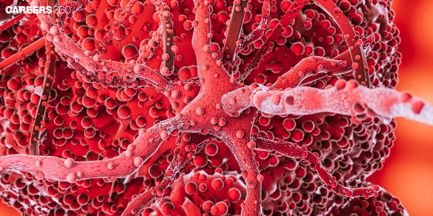
Blood coagulation is the process through which it becomes thicker to form a clot stopping blood from coming out when there is an injury. This process involves several steps and special proteins in blood so that the body does not lose too much blood. This topic is included in the class 11 chapter body fluids and circulation. Questions from this topic are asked in competitive exams like NEET where biology is one of the main subjects.
NEET 2025: Mock Test Series | Syllabus | High Scoring Topics | PYQs
NEET Important PYQ's Subject wise: Physics | Chemistry | Biology
New: Meet Careers360 B.Tech/NEET Experts in your City | Book your Seat now
- What is Blood Coagulation?
- Blood Coagulation Process
- How Do Coagulation Factors Work?
- Side Effects of Coagulation Factors
- Clotting Time
- What is Deep Vein Thrombosis?
- Blood Coagulation Factors
- Disorders of Blood Coagulation
- Recommended Video on Blood Coagulation

What is Blood Coagulation?
Blood coagulation or clotting is a complicated process in which blood is changed from liquid to gel, forming a blood clot to prevent maximum blood loss due to damage to the vessel. This vital function comprises a sequence of events through which the activation of platelets and plasma proteins takes place to form a stable fibrin clot.
Read More:
What is Coagulation?
Coagulation is a crucial phase in the healing process of a wound when it functions as it should. Coagulation aids in the formation of a clot, which is made of a material called fibrin, when a blood vessel breaks, as with a cut or other damage. Until the tissues can heal themselves, the clot closes the wound.
Blood Coagulation Process
The blood coagulation process represents a well-orchestrated sequence involving rapid and effective clotting. All these stages include vascular spasm, platelet plug formation, the coagulation cascade, clot retraction and repair, and finally fibrinolysis.
Vascular Spasm
Vascular spasm refers to the instant contraction of blood vessels upon injury.
Mechanism of Action: Smooth muscles in the blood vessel wall contract, reducing blood flow.
In Blood Coagulation: It provides a temporary seal minimising blood loss.
Formation Of Platelet Plug
The adherence of platelets to the site of injury and their subsequent aggregation by each other onto fibrinogen, thereby forming a temporary "plug".
Platelet Plug Formation Stages
Adhesion: The platelets are associated with exposed collagen fibres of the injured vessel.
Activation: Chemicals released by the platelets attract more platelets.
Aggregation: Platelets clump together, forming the plug.
Platelets thus play a twin role in forming the plug and in initiating the coagulation cascade.
How Do Coagulation Factors Work?
Drugs called coagulation factor concentrates are used to treat haemophilia and control bleeding. A severe impairment of blood clotting caused by haemophilia causes considerable blood loss in the event of even a minor wound. Usually, it results from a hereditary deficiency in a coagulation factor, most frequently factor VIII. Blood-clotting proteins called coagulation factors are produced naturally in the human body. The body's many coagulation components combine to generate clots. The clots stop the body from losing too much blood.
In order for the blood to adequately clot, the missing blood clotting factor must be replaced during haemophilia treatment. To stop serious blood loss, coagulation factor concentrates take the place of the blood clotting factor and help the blood clot.
Side Effects of Coagulation Factors
When injected, coagulation factors may have the following adverse effects:
Dyspnea (shortness of breath)
Fever
Nausea
Dizziness
Headache
Taste disturbance
Itching
Rash
Swelling or redness at the injection site
Stuffy nose.
Coagulation Cascade
Enzymatic cascade leads to the formation of fibrin strands that stabilise the platelet plug.
Coagulation Cascade Pathways
Intrinsic Pathway: This pathway is triggered by an injury in the vascular wall.
Extrinsic Pathway: It gets activated on trauma, and blood is lost from the lumen of the vessel.
Common Pathway: The intrinsic and extrinsic pathways merge to yield the production of fibrin.
The involved factors range from I to XIII and include thrombin-like enzymes.
Clot Retraction and Repair
Shrinking of the clot itself to reduce its size, drawing the edges of the wound closer together.
Mechanism of Clot Retraction: Platelets contract, pulling on fibrin strands.
Process for Tissue Repair and Generation: Tissue repair mechanisms are activated.
Fibrinolysis
The breakdown of the clot after the vessel has healed itself.
Plasminogen activates plasmin, which digests fibrin.
Plasmin is the major proteolytic enzyme responsible for clot dissolution.
Clotting Time
Clotting timings gauge how long it takes for a clot to develop. In the majority of tests, an activator is utilised to start coagulation and assess how well one or more parts of the coagulation cascade model respond. A reduced number or function of the coagulation components involved might cause abnormalities in clotting times.
Prothrombin time (PT) and activated partial thromboplastin time are the two clotting time measurements that are most frequently utilised (aPTT).
Most clinical pathology laboratories offer these tests, which are often carried out by automated coagulation analysers.
Precautions for Clotting Time
Mild coagulation abnormalities are not diagnosed.
Blood should be drawn as gently as possible.
To obtain a precise result, one must prevent premature activation of the clotting process.
Prevent the sample from hemolyzing.
It is crucial to learn the patient's history.
Drawbacks of Clotting Time
This test is unreliable for identifying bleeding conditions.
To identify slight bleeding problems, it is insensitive.
Only the most serious bleeding conditions will be detected.
The presence of aberrant coagulation cannot be ruled out by normal clotting time.
The test's execution involves numerous variables.
For regular preoperative screening for bleeding, such as in tonsillectomy, preoperative clotting time and bleeding time are of minimal benefit.
The Procedure of Clotting Time
Three methods can be estimated for clotting time:
Capillary Method
Use the lancet to prick the finger.
The capillary will instantly fill if you place it above the blood.
Now, rupture the capillary at regular intervals.
The endpoint and clotting time is when a clot begins to form.
Test Tube Method
Conduct this experiment at 37 ° C.
Start the timer after collecting 4 ml of blood with the tube method. Take note of the moment the clot development initially appears.
To improve accuracy, this test can be performed in many tubes.
Lee and White Method
Take two 10-centimetre externally borated siliconised tubes.
These tubes are prewarmed in a water bath at 37 °C.
Take a sample of blood, primarily from the antecubital vein.
1 mL of the blood is placed in each test tube after 2 to 2.5 mL of the blood is drawn.
As soon as you notice the blood in the syringe, start two stopwatches.
Keep the blood in the water bath and tilt each tube every 30 to 60 seconds to check for clotting.
Tilt the tube beyond a 90-degree angle.
As soon as you notice the clot in the tube, stop the stopwatch.
Disadvantages of Clotting Time
This test has lost its value because it is insensitive.
The method used to administer the test involves a lot of variables.
This is unable to identify a mild coagulation factor deficit.
This test is extended only in cases of extreme deficit.
Even while thrombocytopenia causes longer bleeding times, normal clotting times still exist.
What is Deep Vein Thrombosis?
Deep vein thrombosis or DVT refers to a circulatory disorder characterised by the formation of a blood clot or thrombus in the deep vein, usually of the lower limbs.
Causes: Prolonged immobility, surgery, certain medications.
Symptoms: Swelling, pain, redness, and warmth in the affected leg.
Complications: The clot breaks loose and travels in the bloodstream to the lungs where it causes a pulmonary embolism.
What is Thrombus or Thrombosis?
A blood clot in the circulatory system is known as a thrombus. It adheres to the area where it originated and stays there, obstructing blood flow. The medical word for the development of a thrombus is thrombosis.
A clot developing inside a blood vessel (an artery or vein) in your body, or occasionally inside your heart, is called thrombosis. This is a risk because blood vessel clots can restrict blood flow. They can also separate and move about throughout your body, and if a clot lodges itself in a vital organ like your brain or lungs, it may result in emergencies that could endanger your life.
People who are sedentary and those who have a hereditary propensity to blood clotting are more likely to develop thrombi. A thrombus can also develop after an artery, vein, or surrounding tissue is damaged.
Blood Coagulation Factors
Various factors could either impair or enhance blood coagulation, some of which will be discussed including specific proteins, genes, and lifestyle choices.
Coagulation Factors
Plasma proteins that are essential for blood clotting.
Major coagulation factors: I to XIII; each factor has a different function in the coagulation cascade, like Factor VIII in haemophilia A.
Genetic Factors
Hemophilia: Genetic deficiency of clotting factors (Factor VIII or IX).
Symptoms: Excessive bleeding, easy bruising.
Treatment: Replacement therapy with clotting factor concentrates.
Von Willebrand Disease: Deficiency or dysfunction of von Willebrand factor.
Symptoms: Nosebleeds, heavy menstrual periods.
Treatment: Medications are used to increase the von Willebrand factor or clotting factor concentrates.
Environmental and Lifestyle Factors
Diet and Nutrition: Vitamin K is needed for the synthesis of clotting factors.
Medications and Blood Thinners: Aspirin, warfarin, and another anticoagulant will interfere with coagulation.
Exercise and Physical Activity: Regular activity improves circulation, and prevents clot formation.
Causes of Thrombosis
The first step in preventing thrombosis is to determine the likelihood that it will occur. Some persons are more susceptible to thrombosis and the potential for thromboembolism.
The three elements required for the development of thrombosis—blood stasis, vascular wall damage, and altered blood coagulation—have been referred to as "Virchow's triad."
While some risk factors enhance the likelihood of arterial thrombosis, others predispose to venous thrombosis. The risk of thromboembolism in newborn infants during the neonatal era is also present.
Prevention of Thrombosis
Heparin is frequently used after surgery if there are no bleeding complications. A risk-benefit analysis is typically necessary because all anticoagulants raise the risk of bleeding.
Disorders of Blood Coagulation
The disorders lead to either an increase or a decrease in clotting, both conditions presenting significant health risks.
Hypercoagulability
Excess clotting tendency due to genetic factors, drugs, or conditions such as cancer.
Associated Conditions of Hypercoagulability: Deep vein thrombosis, pulmonary embolism
Treatment Options and Management: Anticoagulants, lifestyle modifications
Hypercoagulability
The low ability of blood to form clots is usually due to genetics or as an effect of drugs.
Conditions Associated with Hypocoagulability: Haemophilia, von Willebrand disease.
Treatment and Management: Replacement of clotting factors and supportive therapies.
Blood Coagulation Diagnostic Tests
Several tests are used to diagnose coagulation disorders and monitor the treatment of these disorders.
Prothrombin Time (PT)
Purpose and Procedure: This test will determine how long it takes for blood to clot. It measures the extrinsic pathway.
Interpretation of Results: Prolonged PT indicates clotting disorders or vitamin K deficiency.
Activated Partial Thromboplastin Time
Purpose and Procedure: This is a measure of the time blood takes to clot, and tests intrinsic pathways.
Interpretation of Results: Long aPTT is indicative of haemophilia or heparin therapy.
D-Dimer Test
Purpose and Procedure: Assay fragments produced in the process of clot breakdown; are used to diagnose thrombotic disorders.
Interpretation of Results: Elevated values indicate active clot formation and lysis.
Platelet Count
Purpose and Procedure: Determine the number of platelets present in blood.
Interpretation of Results: Too few, thrombocytopenia, excessive bleeding; too many, thrombocytosis, tendency to form clots.
Also Read:
Recommended Video on Blood Coagulation
Frequently Asked Questions (FAQs)
Vascular spasm, platelet plug formation, coagulation cascade, clot retraction and repair, and fibrinolysis.
They bind to the damaged site; then, by releasing chemicals, they attract more platelets and act as a temporary plug.
The intrinsic pathway begins with internal damage to blood vessels and contains all the factors that are part of the blood. In contrast, the extrinsic pathway begins from external trauma, which includes tissue factors outside the blood.
Common disorders include haemophilia, von Willebrand disease, deep vein thrombosis, and pulmonary embolism.
Diagnosed using tests such as Prothrombin Time, Activated Partial Thromboplastin Time, D-Dimer test, and platelet count.
Blood coagulation is the process by which blood forms a clot to stop bleeding when a blood vessel is injured. It involves the activation of proteins called clotting factors, leading to the formation of a fibrin mesh that seals the wound and prevents further blood loss.
Also Read
29 Nov'24 01:19 PM
27 Nov'24 07:39 PM
27 Nov'24 07:15 PM
27 Nov'24 05:11 PM
26 Nov'24 08:14 PM
26 Nov'24 06:50 PM
26 Nov'24 05:51 PM
26 Nov'24 04:44 PM
26 Nov'24 03:52 PM
26 Nov'24 02:55 PM