Composition of Bacterial Cell Wall: Overview, Topics, Structure
The bacterial cell wall is made up of peptidoglycan, a complex molecule of sugars and amino acids. The composition of the bacterial cell wall varies between bacteria, with gram-positive bacteria having a thick peptidoglycan layer, while gram-negative bacteria have a thinner layer and an outer membrane. The bacterial cell wall structure is essential for maintaining cell shape and protecting the cell. A bacterial cell wall diagram typically shows the differences between gram-positive and gram-negative bacteria. This is an important topic of biology as it links some of the major chapters.
NEET 2025: Mock Test Series | Syllabus | High Scoring Topics | PYQs
NEET Important PYQ's Subject wise: Physics | Chemistry | Biology
New: Meet Careers360 B.Tech/NEET Experts in your City | Book your Seat now
The bacterial cell wall is a firm, protective layer outside the cell membrane, essential for maintaining the bacterium's shape and providing defence against environmental stress. The composition of the bacterial cell wall is primarily made up of peptidoglycan, a polymer that provides strength to the structure.
Gram-positive bacteria have a thick peptidoglycan layer and teichoic acids, while gram-negative bacteria feature a thinner peptidoglycan layer, an outer membrane, and lipopolysaccharides that contribute to antibiotic resistance.
The cell wall structure of gram-positive bacteria is more rigid, as seen in a bacterial cell wall diagram. Overall, the cell wall of bacteria is made up of peptidoglycan and other components that vary between bacterial types. In the end, bacteria have cell walls that are crucial for survival and protection.
Also Read:
Composition of Bacterial Cell Wall
The peptidoglycan a mesh-like polymer of bacterial cell wall provides important rigidity and strength. Teichoic acids are found in Gram-positive bacteria and provide structural stability and function to the cell wall. Lipopolysaccharides are found in the outer membrane of Gram-negative bacteria and are very important in protection from environmental threats and antibiotics.
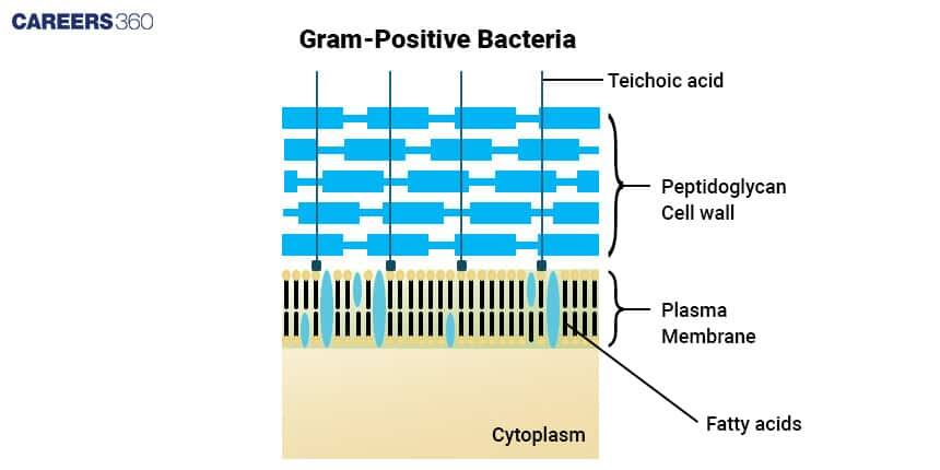
Gram-Positive Bacterial Cell Wall
The bacterial cell wall of gram-positive bacteria is made up of a thick, multilayered peptidoglycan layer, which may constitute up to 90% of the composition of the bacterial cell wall. This extensive peptidoglycan layer provides strength and rigidity to the cell, protecting it against osmotic pressure and mechanical stress.
These acids play important roles in maintaining the bacterial cell wall structure, regulating ions, and contributing to pathogenesis. The cell wall structure of gram-positive bacteria is crucial for their survival, as shown in a bacterial cell wall diagram. Overall, the cell wall of bacteria is made up of peptidoglycan and teichoic acids, ensuring the bacterium's stability.
A diagram of the Gram-positive bacterial cell wall
Gram-Negative Bacterial Cell Wall
The Gram-negative bacterial cell wall is made up of a thin layer of peptidoglycan, which is sandwiched between an inner cytoplasmic membrane and an outer membrane. This outer membrane contains high quantities of lipopolysaccharides, which contribute significantly to the structural integrity of the Gram-negative cell wall and act as a barrier against unwanted access, including that of antibiotics.
The composition of the bacterial cell wall in gram-negative bacteria provides them with greater resistance to environmental stress and antimicrobial agents compared to gram-positive bacteria. This bacterial cell wall structure allows gram-negative bacteria to be more resilient, as shown in a bacterial cell wall diagram. Overall, the cell wall of bacteria is made up of peptidoglycan, lipopolysaccharides, and other components that protect the bacteria.
Defective Bacterial Cell Walls
The bacterial cell wall can become defective due to interventions like antibiotics, lysozymes, and bacteriophages, which block the normal processes of cell wall formation. A bacterial variant without a classical cell wall is called an L-form, which appears after exposure to antibiotics like penicillin.
Mycoplasma is a naturally occurring form of bacteria that lacks a cell wall. Despite their absence of a bacterial cell wall, they are considered a separate group due to their unique characteristics.
Spheroplasts are typically non-recoverable from gram-positive bacteria but can be formed from gram-negative bacteria, whereas protoplasts, which are formed from gram-positive bacteria, are recoverable and viable. These form the ability of bacteria to adapt to and survive environmental and antimicrobial pressures, reflecting their ability to modify their bacterial cell wall structure.
Also Read
Frequently Asked Questions (FAQs)
Gram-positive bacteria contain thick peptidoglycan and teichoic acids, while Gram-negative bacteria have thin peptidoglycan sandwiched between an inner and outer membrane; most of the latter contain lipopolysaccharides.
These penicillins are acting on the transpeptidase enzyme, which normally cross-links the peptidoglycan strands. These ultimately result in the weakening of the cell wall, and the foremost consequence is the lysis of bacterial cells.
This provides the bacterial cell wall with rigidity and structural support that protects the cell from osmotic pressure and helps in the shape maintenance of the cell.
Gram-negative bacteria have an outer membrane barrier that prevents many antibiotics from reaching their target sites within the cell wall or membrane.
Bacterial adhesion, colonisation, and evasion of host immune responses are facilitated by a component such as lipopolysaccharides and teichoic acids, thereby enhancing the ability to cause disease.
Also Read
30 Nov'24 12:23 PM
28 Nov'24 05:34 PM
25 Nov'24 05:18 PM
23 Nov'24 10:02 AM
22 Nov'24 01:59 PM
21 Nov'24 04:58 PM
16 Nov'24 01:58 PM