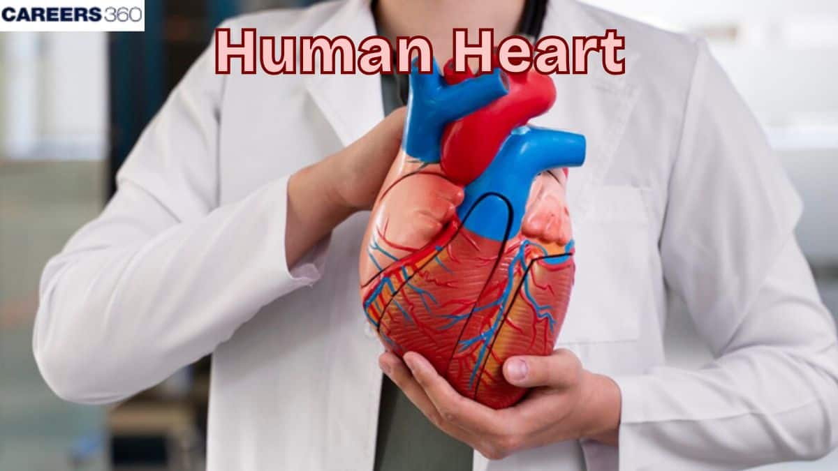
Human Heart

Human Heart

Edited By Irshad Anwar | Updated on Jul 02, 2025 06:41 PM IST

The human heart is an important muscular organ that pumps blood. It has four chambers—two atria and two ventricles—that work together to circulate oxygen-rich blood to the different organs. In this article, the human heart, the anatomy of the human heart, functions of the human heart, facts about the human heart, and heart health are discussed. Human heart is a topic of the chapter Body Fluids and Circulation in Biology.

This Story also Contains
  1. Human Heart
  2. Anatomy of the Human Heart
  3. Functions of the Human Heart
  4. Facts About The Human Heart
  5. Heart Health
Human Heart
Human Heart

Human Heart

The human heart is one of the human organs providing vital support to the flow of blood in the human body through the circulation of blood to deliver new oxygen, and nutrients to the tissues and organs; in return, it removes the waste products from these tissues and organs. It is basic to understand the function and significance of the heart considering that it pumps blood and keeps the blood circulation within acceptable limits. Structurally, the heart has four sections; two upper chambers or the atrium and two lower chambers known as the ventricles; these valves control the direction of blood circulation. These contractile movements are due to the creation of muscular walls, and commands of electrical impulses also help the heart to be a perfect pump that will supply the health and proper functioning of the entire body system.

Anatomy of the Human Heart

The anatomy of the human heart is discussed below-

Location And Size

  • The human heart is situated in the mediastinum, more specifically in the thoracic cavity and in front of the lungs in coordination with the left side of the sternum axis.

  • It is situated behind the sternum and above the diaphragm, although is located in the mediastinum.

  • Specifically, an average human heart is as large as a fist and weighs between 250-350gm (9-12oz) in an adult.

Heart Location in the Body

Heart Structure

The human heart is made of the following components:

Pericardium

  • The overall structure in which the heart is surrounded is called the pericardium and is made up of two elementary structures, namely the fibrous pericardium and the serous pericardium.

  • Serous pericardium, in turn, gets subdivided into the parietal and visceral layers, whereas the latter is referred to as the epicardium.

  • The pericardium acts as a shield to the heart, it holds the heart in position and acts as a constraint to the heart’s ventricular filling.

NEET Highest Scoring Chapters & Topics
Know Most Scoring Concepts in NEET 2024 Based on Previous Year Analysis.
Know More

Heart wall

  • Epicardium: The first one is the protection layer for giving protection to the inner part.

  • Myocardium: The middle, the muscular layer is characterized by the power of contraction and performing the act of pumping.

  • Endocardium: The endocardium is the inner layer that faces the blood flow in the heart chambers and over the valves.

Chambers

  • Atria: The two right and left atria are the upper chambers of the heart that receive the blood coming to the heart.

  • Ventricles: The last two chambers of the heart are the ones that pump blood out of the heart, these are the right and left ventricles.

Valves

The human heart is muscular and relatively small in size, including four valves that ensure one-way blood circulation.

  • Atrioventricular Valves: These two are the tricuspid valve, belonging to the right side of the heart and the mitral valve on the left side of the heart.

  • Semilunar Valves: This includes; the pulmonary valve at the right side and the aortic valve at the left side.

Internal Structure of the Heart

Blood Vessels

The blood vessels of the heart include:

Arteries

  • Aorta: The largest artery; carries oxygenated blood from the left ventricle through the aortic arch and onto the body.

  • Pulmonary Arteries: Transport deoxygenated blood from the right ventricle and take it to the lungs.

Veins

  • Superior and Inferior Vena Cava: Pump deoxygenated blood from the body back to the right atrium.

  • Pulmonary Veins: Pump oxygenated blood from the lungs and deliver it to the left atrium.

Human Heart Diagram

Major Blood Vessels of the Heart

Conduction System

  • SA Node (Sinoatrial Node): The heart also contains a small organ called the pacemaker which is sited in the right atrium to determine the rate of contraction.

  • AV Node (Atrioventricular Node): It is located between the atria and the ventricles; it gets impulses for the stimulation of the contractions from the SA node and conveys them to the ventricles.

  • Bundle of His: An avenue for electrical impulses originating from the AV node to the ventricles.

  • Purkinje Fibers: Bundle of nerves that convey electrical stimulus to other chambers of the heart namely the ventricles to make them contract.

Heart’s conduction system

Also Read-

Functions of the Human Heart

The functions of the human heart are discussed below-

Pumping Blood

The blood in the body is circulated by the heart through two main loops which are known as circuits.

  • Systemic Circuit: Pumps the oxygenated blood from the left ventricle to the different body parts and brings back the deoxygenated blood in the right atrium.

  • Pulmonary Circuit: Brings deoxygenated blood from the right ventricle to the lungs to get oxygenated and pumps oxygenated blood to the left atrium.

Cardiac cycle:

The mechanisms of the cardiac cycle may be divided into two major stages:

  • Systole: The heartbeat cycle, the period that describes the contraction of the heart muscle, followed by pumping blood out of the atrial or ventricular chambers into the arteries.

  • Diastole: The period in which the heart muscle is at rest, thus the chambers receive blood.

Blood Flow through the Heart

Maintaining Blood Pressure

  • Many factors are involved in the regulation of blood pressure; the force and rate of the contractions of the heart.

  • This helps avoid inadequate blood flow in the body to meet the necessary needs of the human body.

  • The circulation system also involves blood vessels which can also narrow or expand to assist in the regulation of blood pressure.

  • More specifically, arteries are in charge of providing rather constant pressure and blood flow in this regard.

Supplying Oxygen And Nutrients

  • Oxygenated blood is developed for the process of respiration where the cells get their required oxygen for the production of energy.

  • The circulatory system transports material such as glucose and amino acids in their developed forms to cells and embraces products such as carbon dioxide and urea.

Endocrine Function

  • It also plays the role of an endocrine gland because it has the capability of secreting hormones that control other activities in the body.

  • Similar to CNP, ANP is released from the atria of the heart when blood volume and pressure are high.

  • ANP aids in the prevention of an increase in blood volume by promoting sodium and water elimination in the urine.

  • ANP has significant effects on controlling the blood pressure and regulating the fluid volume in the body hence it is involved in the body’s homeostasis.

Circulation Types

  • A part of circulation called pulmonary circulation is in charge of returning oxygenated blood to the heart after transporting deoxygenated blood to the lungs.

  • Another aspect of circulation is systemic circulation, in which the heart pumps oxygenated blood to all of the body's organs and tissues while returning deoxygenated blood to the heart.

  • Coronary Circulation includes coronary arteries that supply oxygenated blood to the myocardium; and coronary veins that carry deoxygenated blood away from the heart muscle.

Facts About The Human Heart

  • The human heart rate is determined by the number of times it beats per minute; this ranges from 60-100 bpm when a person is at rest.

  • Pulse can be felt by lightly palpating arteries underneath the skin specifically the radial pulse at the wrist or the carotid pulse on the neck.

  • Finding out the total number of beats in one minute will indicate the rate at which the heart is beating.

  • Cardiac output is defined as the volume of blood that is pumped by the heart within one minute.

  • It plays a very important role in the provision of oxygen and nutrients to all body tissues.

  • Cardiac output is affected by physical activity, the size of the body and the overall health of a person.

Heart Health

Caring for our hearts is among the core values for the quality of life free from diseases, mishaps, and untimely deaths.

Coronary Artery Disease

A condition that affects the blood vessels called the coronary arteries leading into the heart muscle and which get blocked thus limiting the amount of blood that is delivered to the muscle.

Heart Attack

This happens when a section of the heart does not get an adequate amount of blood and nutrients hence becoming damaged.

Heart Failure

A state in which the heart is not able to pump blood as it should, hence accumulating fluids within the body.

Preventive measures:

  • Diet: A healthy diet characterised by increased consumption of fruits and vegetables, whole grain products, lean meats, and healthy fats helps to maintain healthy bones and muscles.

  • Exercise: Physical activity in postmenopausal women has been established to be beneficial as it enhances the working capacity of the heart muscle, and circulation and keeps the body lean.

  • Lifestyle Changes: Not smoking, taking a moderate amount of alcohol, learning how to handle stress and getting checkups are important in the promotion of heart health.

Also Read-

Recommended Video for Human Heart


Frequently Asked Questions (FAQs)

1. What are the main functions of the human heart?

This muscle efficiently sends oxygenated blood to the body and deoxygenated blood through the pulmonary artery and/or sends blood pressure and nutrients and removes waste products from the body. 

2. What are the different parts of the heart?

While the heart has two atria – right and left, and two ventricles – right and left; the valves include tricuspid, pulmonary, mitral and aortic; layers of the heart walls comprise epicardium, myocardium, and endocardium. Some of the important arteries and veins include the aorta, arteries through the lungs, the veins through the lungs, and the vena cava.

3. How does the heart's conduction system work?

The SA node initiates the heartbeat, the AV node passes the impulse, and the Bundle of His conducts the impulse till the ventricles and Purkinje fibres distribute the impulse to the Ventricle to contract.

4. What are common heart diseases and how can they be prevented?

Some of the diseases include; coronary artery disease, myocardial infarction also known as heart attack, and heart failure. There are vigorous measures that an individual and or a patient can take such include observing a balanced diet, exercising daily, refraining from smoking, moderating instances of alcohol intake and ways of handling stress among others.

5. How can I maintain a healthy heart?

Adopt beneficial habits by taking foods in their right proportions, engaging in physical activities, not using tobacco and taking moderate alcohol, managing stress and getting appropriate medical examinations.

6. How many chambers does the human heart have, and what are their names?
The human heart has four chambers: two upper chambers called atria (singular: atrium) and two lower chambers called ventricles. The right atrium and right ventricle form the right side of the heart, while the left atrium and left ventricle form the left side. These chambers work together to pump blood through the pulmonary and systemic circulations.
7. What is the significance of the heart's apex?
The heart's apex is the pointed lower end of the heart, formed mainly by the left ventricle. It's significant because it's the part of the heart closest to the chest wall, where the heartbeat can be most easily felt or heard. The apex's position and movement during the cardiac cycle provide important diagnostic information about heart function and can be assessed through physical examination or imaging techniques.
8. What is the role of coronary arteries in heart function?
Coronary arteries supply oxygen-rich blood to the heart muscle itself. They branch off from the aorta and run along the surface of the heart, delivering vital oxygen and nutrients to cardiac tissue. This blood supply is crucial for the heart's continuous pumping action. Blockage or narrowing of these arteries can lead to reduced blood flow to the heart muscle, potentially causing a heart attack.
9. What is the difference between arteries and veins in relation to heart function?
Arteries carry blood away from the heart under high pressure, while veins return blood to the heart under low pressure. Arteries have thick, elastic walls to withstand the pressure of blood ejected from the heart. Veins have thinner walls and contain valves to prevent backflow of blood as it moves against gravity. Understanding this difference is crucial for comprehending the overall circulatory system and heart function.
10. How does the pericardium contribute to heart function?
The pericardium is a double-layered sac that surrounds the heart. It serves several important functions: it anchors the heart within the chest cavity, provides a friction-reducing surface for heart movement, prevents overfilling of the heart with blood, and acts as a barrier against infection. The fluid between the pericardial layers also helps distribute the forces of heart contraction, contributing to efficient pumping action.
11. How does the fetal heart differ from an adult heart?
The fetal heart differs from an adult heart in several ways. It has additional structures like the foramen ovale (an opening between atria) and the ductus arteriosus (a connection between the pulmonary artery and aorta). These allow blood to bypass the non-functioning fetal lungs. The fetal heart also pumps a higher percentage of its output to the upper body and brain. After birth, these fetal structures close, and the circulation pattern changes to the adult form.
12. How does the autonomic nervous system regulate heart function?
The autonomic nervous system regulates heart function through its two branches: sympathetic and parasympathetic. The sympathetic nervous system increases heart rate and contractility, preparing the body for "fight or flight" responses. The parasympathetic system, primarily via the vagus nerve, slows heart rate and promotes relaxation. This dual control allows for fine-tuning of heart function in response to various physiological demands and environmental conditions.
13. How does the heart maintain its own blood supply?
The heart maintains its own blood supply through the coronary circulation. Coronary arteries, which branch off from the aorta just above the aortic valve, supply oxygen-rich blood to the heart muscle. The left coronary artery supplies the left side of the heart, while the right coronary artery supplies the right side and back of the heart. This self-supply system is crucial because the heart muscle requires a constant supply of oxygen and nutrients to function properly.
14. What is the significance of heart rate variability?
Heart rate variability (HRV) refers to the variation in time intervals between heartbeats. It's an indicator of the heart's ability to respond to various physiological and environmental stimuli. High HRV generally indicates good cardiovascular health and a well-functioning autonomic nervous system. Low HRV can be associated with increased stress, poor cardiovascular health, or certain medical conditions. Understanding HRV provides insights into overall heart health and autonomic balance.
15. What is the role of atrial natriuretic peptide (ANP) in heart function?
Atrial natriuretic peptide (ANP) is a hormone produced by heart muscle cells in the atria. It's released in response to stretching of the atrial walls, typically due to increased blood volume. ANP plays a crucial role in regulating blood pressure and volume by promoting sodium and water excretion in the kidneys, relaxing blood vessels, and inhibiting the renin-angiotensin-aldosterone system. This hormone exemplifies how the heart functions not just as a pump, but also as an endocrine organ.
16. What is the primary function of the human heart?
The primary function of the human heart is to pump blood throughout the body. It acts as a muscular pump that circulates oxygen-rich blood to tissues and organs, while also collecting deoxygenated blood to be reoxygenated in the lungs. This continuous circulation is essential for delivering nutrients, removing waste products, and maintaining overall bodily functions.
17. What is the difference between the left and right sides of the heart?
The left and right sides of the heart have different functions. The right side pumps deoxygenated blood to the lungs for oxygenation (pulmonary circulation), while the left side pumps oxygenated blood to the rest of the body (systemic circulation). The left side has thicker muscular walls to generate higher pressure needed to pump blood throughout the entire body.
18. What are heart valves, and how do they function?
Heart valves are flap-like structures that ensure blood flows in the correct direction through the heart. There are four main valves: tricuspid, pulmonary, mitral, and aortic. They open to allow blood flow and close to prevent backflow. This one-way flow mechanism is crucial for efficient blood circulation and preventing blood from flowing backward within the heart chambers.
19. How does the heart's electrical conduction system work?
The heart's electrical conduction system coordinates the contraction of heart muscles. It begins with the sinoatrial (SA) node, often called the heart's natural pacemaker, which generates electrical impulses. These impulses spread through the atria, then to the atrioventricular (AV) node, and finally through the bundle of His and Purkinje fibers to the ventricles. This system ensures the heart chambers contract in the correct sequence for efficient pumping.
20. What is the cardiac cycle, and what are its main phases?
The cardiac cycle refers to the sequence of events that occur during one heartbeat. It consists of two main phases: systole (contraction) and diastole (relaxation). During systole, the ventricles contract to pump blood out of the heart. During diastole, the ventricles relax and fill with blood. This cycle repeats continuously, typically 60-100 times per minute in a resting adult.
21. How does the heart adapt to increased physical activity?
During physical activity, the heart adapts by increasing both its rate (beats per minute) and stroke volume (amount of blood pumped per beat). This results in increased cardiac output, delivering more oxygen-rich blood to working muscles. The autonomic nervous system, particularly the sympathetic nervous system, plays a key role in this adaptation by stimulating the SA node and increasing heart contractility.
22. How does blood pressure relate to heart function?
Blood pressure is the force exerted by blood against the walls of arteries. It's typically measured as systolic (during heart contraction) and diastolic (during heart relaxation) pressures. The heart's pumping action creates this pressure, which is necessary to circulate blood throughout the body. Maintaining proper blood pressure is crucial for heart health and overall cardiovascular function.
23. What is cardiac output, and how is it calculated?
Cardiac output is the volume of blood pumped by the heart in one minute. It's calculated by multiplying heart rate (beats per minute) by stroke volume (amount of blood pumped per beat). For example, if the heart rate is 70 beats per minute and stroke volume is 70 mL, cardiac output would be 4900 mL or about 5 liters per minute. This measure is crucial for assessing overall heart function and circulatory efficiency.
24. How do heart sounds relate to the cardiac cycle?
Heart sounds, often described as "lub-dub," correspond to specific events in the cardiac cycle. The first sound ("lub") occurs when the mitral and tricuspid valves close at the beginning of systole. The second sound ("dub") is produced when the aortic and pulmonary valves close at the end of systole. Listening to these sounds (auscultation) can provide valuable information about heart valve function and overall cardiac health.
25. How does the Frank-Starling mechanism regulate heart function?
The Frank-Starling mechanism is a physiological principle stating that the heart will pump out whatever amount of blood it receives (within physiological limits). As more blood flows into the ventricles, the cardiac muscle fibers stretch more, leading to a stronger contraction and increased stroke volume. This intrinsic mechanism allows the heart to adapt to changing blood flow demands without nervous system input, ensuring efficient circulation.
26. What is the role of the interventricular septum?
The interventricular septum is the muscular wall that separates the left and right ventricles of the heart. It plays a crucial role in maintaining the separation of oxygenated and deoxygenated blood. The septum also contributes to the heart's pumping efficiency by providing structural support and participating in ventricular contraction. Defects in this septum can lead to mixing of oxygenated and deoxygenated blood, causing serious circulatory problems.
27. How does the positioning of the heart in the chest cavity affect its function?
The heart is positioned in the mediastinum, slightly to the left of the midline in the chest cavity. This positioning allows for efficient blood distribution to all parts of the body. The heart's orientation, with the apex pointing down and to the left, influences the direction of blood flow and the effectiveness of its pumping action. The surrounding structures, including the lungs and diaphragm, also play a role in supporting heart function and influencing its movement during the cardiac cycle.
28. How does the heart's double-pump system contribute to efficient circulation?
The heart functions as a double pump, with the right side pumping deoxygenated blood to the lungs (pulmonary circulation) and the left side pumping oxygenated blood to the body (systemic circulation). This separation ensures that oxygenated and deoxygenated blood don't mix, maximizing oxygen delivery efficiency. The double-pump system also allows for different pressures in the pulmonary and systemic circulations, optimizing gas exchange in the lungs and blood flow to body tissues.
29. What is the role of gap junctions in heart muscle function?
Gap junctions are specialized membrane channels that directly connect the cytoplasm of adjacent cardiac muscle cells. They allow for the rapid passage of electrical impulses between cells, enabling the heart muscle to act as a functional syncytium (a mass of cytoplasm with multiple nuclei). This electrical coupling ensures synchronized contraction of heart muscle cells, which is crucial for efficient pumping. Understanding gap junctions is key to grasping how the heart achieves coordinated contraction across its chambers.
30. What is cardiac reserve, and why is it important?
Cardiac reserve refers to the heart's ability to increase its output above resting levels during periods of increased demand, such as during exercise. It's the difference between the maximum cardiac output and the resting cardiac output. A healthy heart has a significant cardiac reserve, allowing it to meet increased metabolic demands without undue stress. This concept is important for understanding how the heart adapts to various physiological states and for assessing overall cardiovascular health.
31. What is cardiac hypertrophy, and how does it affect heart function?
Cardiac hypertrophy is an increase in the size of heart muscle cells, leading to thickening of the heart walls. It can be a normal adaptation to increased workload (as in athletes) or a pathological condition (as in hypertension or heart disease). While initially compensatory, excessive hypertrophy can lead to reduced cardiac efficiency, increased oxygen demand, and potential heart failure. Understanding this concept is crucial for differentiating between physiological and pathological heart changes.
32. What is the role of the bundle of His in heart function?
The bundle of His is a specialized group of cardiac fibers that conducts electrical impulses from the atrioventricular (AV) node to the ventricles. It divides into left and right bundle branches, which further divide into Purkinje fibers. This conduction system ensures that the electrical signal reaches all parts of the ventricles almost simultaneously, allowing for coordinated and efficient ventricular contraction. Disruptions in this system can lead to various types of heart block and arrhythmias.
33. What is the significance of the Purkinje fibers in heart function?
Purkinje fibers are specialized cardiac muscle fibers located in the inner ventricular walls. They are the final part of the heart's electrical conduction system. These fibers conduct electrical impulses much faster than regular cardiac muscle, ensuring that the ventricles contract quickly and in a coordinated manner. This rapid and synchronized contraction is crucial for efficient blood pumping. Understanding the role of Purkinje fibers is essential for comprehending how the heart achieves its coordinated pumping action.
34. How does the heart adapt to changes in body position?
The heart adapts to changes in body position through baroreceptor reflexes and the Frank-Starling mechanism. When moving from lying to standing, for example, blood tends to pool in the lower body due to gravity. Baroreceptors detect the resulting drop in blood pressure, triggering an increase in heart rate and contractility to maintain adequate blood flow to the brain. The Frank-Starling mechanism also helps by increasing stroke volume as more blood returns to the heart when lying down.
35. How does the thickness of heart chamber walls relate to their function?
The thickness of heart chamber walls is directly related to their function and the pressure they must generate. The left ventricle, which pumps blood to the entire body, has the thickest walls to generate high pressure. The right ventricle, pumping only to the lungs, has thinner walls. Atria have the thinnest walls as they mainly receive blood and contract against little pressure. This structural adaptation ensures each chamber can perform its specific role efficiently in the overall circulation process.
36. What is the role of the moderator band in heart anatomy and function?
The moderator band, also known as the septomarginal trabecula, is a band of cardiac muscle in the right ventricle. It connects the interventricular septum to the anterior papillary muscle. Its primary function is to carry part of the right bundle branch of the heart's conduction system to the anterior papillary muscle. This ensures that the papillary muscle contracts in coordination with the rest of the ventricle, aiding in proper closure of the tricuspid valve during systole.
37. How does the heart maintain its rhythm without external nervous input?
The heart maintains its rhythm through its intrinsic conduction system, primarily the sinoatrial (SA) node. The SA node, located in the right atrium, contains specialized pacemaker cells that spontaneously generate electrical impulses. These cells have the unique ability to depolarize on their own, creating a regular rhythm. While the autonomic nervous system can modulate this rhythm, the heart can continue beating even without external nervous input, demonstrating its remarkable self-regulatory capacity.
38. What is the significance of the coronary sinus in heart function?
The coronary sinus is a large vein on the posterior surface of the heart that collects deoxygenated blood from the heart muscle itself. It plays a crucial role in the venous drainage of the heart, collecting blood from smaller cardiac veins and emptying into the right atrium. Understanding the coronary sinus is important for comprehending how the heart tissue itself is drained of deoxygenated blood, completing the cycle of the heart's own blood supply and drainage system.
39. How does the heart's energy metabolism differ from other organs?
The heart has unique energy metabolism due to its constant workload. It primarily uses fatty acids for energy, unlike most other organs that prefer glucose. The heart can also use ketone bodies, lactate, and glucose as fuel sources. It has a high density of mitochondria to support its high energy demand. This metabolic flexibility allows the heart to maintain its function under various conditions, including fasting or intense exercise, highlighting its adaptability to different physiological states.
40. What is the role of the chordae tendineae in heart function?
The chordae tendineae, often called "heart strings," are fibrous cords that connect the papillary muscles to the leaflets of the mitral and tricuspid valves. Their primary function is to prevent these valves from prolapsing (bulging back) into the atria during ventricular contraction. By anchoring the valve leaflets, the chordae tendineae ensure proper valve closure, preventing backflow of blood and maintaining the efficiency of the heart's pumping action.
Human Heart

02 Jul'25 06:41 PM

Articles

Back to top