Structure of the Human Eye: Definition, Anatomy, Diagram, Function, & Facts
The human eye is an excessively complex and extremely good organ that lets people look at the sector with first-rate detail. It is essential to most everyday activities, together with reading, using, or maybe facial identity hence, its presence within the human frame is alternatively critical. This is important part of Chapter Neural Control and Coordination in Biology.
NEET 2025: Mock Test Series | Syllabus | High Scoring Topics | PYQs
NEET Important PYQ's Subject wise: Physics | Chemistry | Biology
New: Meet Careers360 B.Tech/NEET Experts in your City | Book your Seat now

Anatomy of the Human Eye
The human eye is such a complex organ, with its structure and function being of high importance for vision. Many parts make up the anatomy of the eye, each contributing to the capture and processing of light to create visual images.
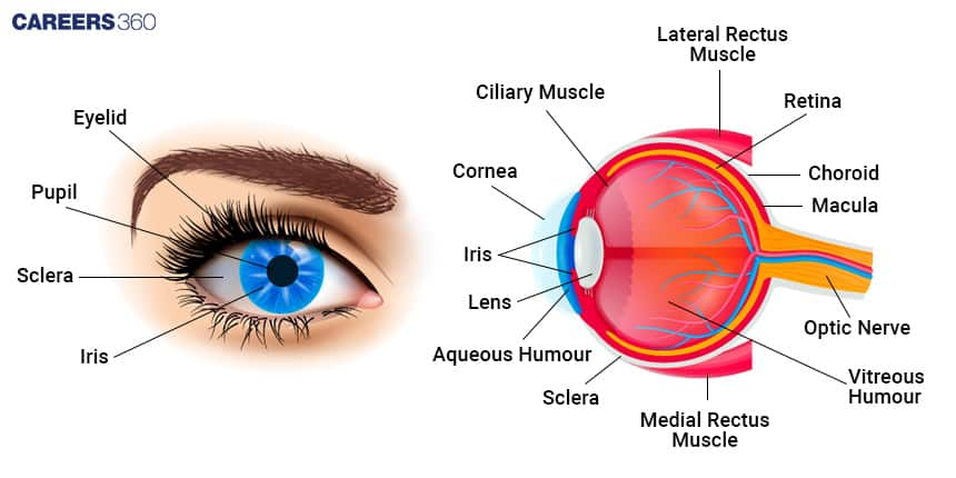
The human eye, as depicted in a human eye diagram, consists of a cornea, lens, retina, iris, and optic nerve. The internal structure of the eye is described with the help of a diagram showing labelling of the parts of the human eye including aqueous humor, vitreous humor, and ciliary muscles.
Eye parts and functions work in unison are the cornea focuses light, the lens adjusts focus, and the retina converts light into nerve signals transmitted to the brain. A simple eye diagram, such as that for class 10, helps explain the structure of the human eye and how it supports vision, with the functions of the eye parts ensuring precise and coordinated visual perception.
Also Read:
External Anatomy
Eyelids: They help to protect the eyes from dust and other extraneous things and spread the tears.
Eyelashes: They collect dust and other minute particles to prevent their falling into the eye.
Sclera: That part of the eyeball that is white, structural, and protective.
Cornea: Transparent anterior portion of the eyeball that helps in focusing light.
Conjunctiva: Thin, semi-transparent membrane covering the sclera and internal eyelids lubricates the attention.
Internal Structure
Anterior Segment:
Cornea: The clear, bell-shaped hood in front of the eye.
Aqueous Humor: It is a type of Fluid present between the cornea and lens.
Iris: This is the coloured part of the eye that changes in size of the pupil.
Pupil: The Center of the iris controlling the quantity of light that may enter.
Lens: The clear structure behind the iris focuses light onto the retina.
Posterior Segment:
Vitreous Humor: The gel-like substance gives shape to the eye.
Retina: the light-sensitive layer on which the images are formed
Macula: The main part of the retina responsible for clear vision
Fovea: It is the depression inside the centre of the macula, providing the clearest imaginative and prescient.
Optic disc: a blind spot in which the optic nerve attaches to the retina.
Supporting Structures:
Choroid: Blood vessel-containing layer, recharging the retina.
Cornea: Outer, protective layer of the eyeball.
Optic Nerve: Transfers visual records from the retina to the mind.

Functions of the Human Eye
Each human eye part performs a distinct role in the functioning of vision.
Cornea
This component's clarity is transparent thus enabling light.
It refracts light that reaches the retina.
Lens
The shape or form changes to focus light into clear vision at the retina.
Distances from near to far are observable.
Retina
Photoreceptor cells, rods and cones.
Light is changed to electrical impulses.
Optic Nerve
The nerve that carries visual impulses from the retina to the brain.
It allows the photo processing and interpretation of the same to be done.
Iris and Pupil
The iris changes its length depending on the amount of light reaching it.
Ensures that excess light does not reach the retina.
Macula and Fovea
Provides sharp, clear central vision.
Used in activities that require reading and driving.
Also Read:
Recommended video on "Structure of the Human Eye"
Frequently Asked Questions (FAQs)
Cornea, lens, retina, iris, pupil, optic nerve, and the sclera.
The human eye works with the refraction of light from the cornea and the lens onto the retina. The photoreceptor cells there convert the light into electric signals which are sent to the brain by way of the optic nerve.
Common eye disorders include cataracts or clouding of the lens, glaucoma, damage to the optic nerve, and macular degeneration or deterioration of the essential retina. These are usually due to growing older, genetics, and way of lifestyle.
A healthy eye care routine would include common eye checkups, UV-included eyeglasses, a balanced weight loss plan supplemented with vitamins A, C and E and accurate eye hygiene.
Some tendencies made inside the area of eye treatment are LASIK surgical treatment of vision correction, Evolved strategies of cataract surgical operation, and Novel remedies for retinal sicknesses, together with gene remedy and retinal implants.
Also Read
30 Nov'24 10:55 AM
29 Nov'24 08:48 PM
29 Nov'24 06:52 PM
29 Nov'24 05:35 PM

