Structure and Functions of Human Eye with Diagram

Structure and Functions of Human Eye with Diagram

Edited By Team Careers360 | Updated on Jul 02, 2025 05:08 PM IST

The eye is one of the body's sense organs. The eye's structure is vital to comprehend because it is one of the most significant sensory organs in the human body. It is primarily responsible for vision, colour discrimination (the human eye can distinguish around 10 - 12 million colours), and the human body's biological clock. The human eye and a camera are similar in that they both gather, focus, and transmit light through a lens to create an image of an object.

Structure and Functions of Human Eye  with Diagram
Structure and Functions of Human Eye with Diagram

The eye's structure is crucial to examine because it is one of the most significant sense organs in humans. The feeling of vision is made possible by the eyes. The human eye's structure is similar to that of a camera. Both of them create images by gathering, focussing, and transmitting light through a lens.

human eye

Also read -

Structure and functions of eye:

The eyeballs are not entirely spherical, contrary to popular perception; instead, they are made up of two different segments fused together.

The human eye is one of the most complex sensory organs in the body. Every element of the human eye, from muscles and tissues to nerves and blood arteries, is accountable for a certain action. Furthermore, contrary to popular assumption, the eye is made up of two different parts fused together, rather than being entirely spherical. It is made up of various muscles and tissues that produce an approximately spherical shape. The human eye can be divided into two categories from an anatomical standpoint: outward structure and internal structure.

Every portion of the human eye has a specific purpose, and these functions are added together to produce the final vision. Internal and exterior structure are the two fundamental components of the eye.

Let's take a look at each one separately.

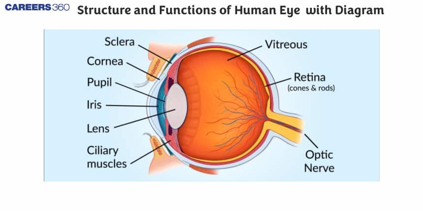
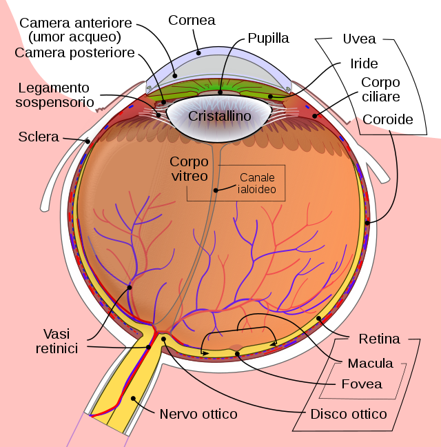
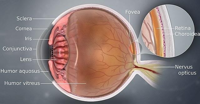
The sclera, conjunctiva, cornea, iris, and pupil make up the exterior structure of the eye. The sclera is the white, dense connective tissue covering on the outside of the eye. Its purpose is to protect the eye's inner workings. The conjunctiva is a layer of stratified squamous epithelium that lies over the sclera. It works to keep our eyes hydrated and clear. Tears and mucus are secreted to offer lubrication.

The cornea is the translucent front portion of the eye. It covers the iris as well as the pupil. Its purpose is to refract light beside the eye's lens. The pigmented region of the eye that determines eye colour is called the iris. It is the middle region of the outer eye that is visible. Its purpose is to keep the pupil's diameter constant in relation to the light source. The pupil is a tiny hole in the centre of the iris that allows light to pass through. It permits light to pass through to the eye. It concentrates the light entering the eye on the retina.

NEET Highest Scoring Chapters & Topics
This ebook serves as a valuable study guide for NEET exams, specifically designed to assist students in light of recent changes and the removal of certain topics from the NEET exam.
Download E-book

The eye's interior structure is a little complicated. The lens, retina, optic nerve, aqueous humour, and vitreous humour are all part of it.

The lens is a clear biconvex structure with connective tissue connecting it to the ciliary body. The lens, in conjunction with the cornea, refracts the light entering the eye and assists in focusing it on the retina. The retina is the most important portion of the eye for vision. It is the eye's deepest layer. It is sensitive to light and looks like camera film. The retina contains color-detecting photoreceptor cells. The image is converted into electrical signals by the retina. The brain then processes these electrical signals. The optic nerve, which is located in the back of the eyes, is in charge of transmitting nerve impulses from the retina to the brain.

The watery fluid that exists between the lens and the cornea is known as aqueous humour. It hydrates the eye while nourishing it. On the other hand, vitreous humour is a clear jelly-like fluid that lies between the retina and the lens. Its purpose is to protect the eye while also maintaining the spherical form of the eye.

Human eye

External structure of eyes:

The following are the portions of the eye that are visible from the outside:-

  1. Sclera is a visible white part of the skin. It protects the inner organs and is made up of dense connective tissue.
  2. The conjunctiva is a layer of stratified squamous epithelium that lines the sclera. It lubricates our eyes and maintains them moist and clear by secreting mucus and tears.
  3. Cornea: The cornea is the transparent, front component of our eye that covers the pupil and iris. The primary purpose of the lens is to refract light.
  4. Iris: The pigmented, coloured region of the eye that is visible from the outside. The iris' primary role is to regulate the diameter of the pupil in response to the light source.
  5. The pupil is the little opening in the centre of the iris. It lets light in and focuses it on the retina.\

Also Read:

Internal structure of eyes:

The following are the internal components of an eye:

  1. The lens of an eye is a clear, biconvex lens. Ligaments connect the lens to the ciliary body. The lens, in conjunction with the cornea, refracts light to concentrate it on the retina.
  2. The retina is the eye's innermost layer. It is light-sensitive and functions as a camera film. There are three layers of neural cells in them: ganglion, bipolar, and photoreceptor cells. It turns a picture into electrical nerve impulses that the brain can perceive.
  3. The ophthalmic nerve is found in the back of the eyes. All nerve impulses from the retina to the human brain are carried via the optic nerves, which are responsible for perception.
  4. Between the cornea and the lens is a watery fluid called aqueous humour. It nourishes and inflates the eyeball.
  5. Vitreous Humour is a jelly-like translucent material found between the lens and the retina. It is made of of 98% water, collage, proteins, and other ingredients. Vitreous humour's primary role is to protect the eyes and maintain their spherical shape.

Also check-

NCERT Physics Notes:

Frequently Asked Questions (FAQs)

1. Explain the structure of the human eye in a few words.

The human eye is a roughly spherical organ that receives visual impulses and processes them. It is contained within the skull's eye sockets and is held in place by muscles within the sockets.


Because the eye is made up of two parts fused together, it does not have a perfectly spherical form. External components are structures visible from the outside of the eye, while internal components are structures found within the eye.


The following are examples of external and internal components:


Retina,Vitreous Humour, aqueous humour ,Sclera ,Conjunctiva ,Cornea, Iris, Pupil.

2. What are the eye's exterior structures?

The following are examples of the eye's exterior structures:


 Sclera, Conjunctiva, Cornea, Iris , Pupil

3. What is the role of the conjunctiva in the body?

The conjunctiva lubricates the eye's front surface. It also keeps debris, dust, and infection-causing bacteria out of the eyes.

4. What is the role of the iris in the human body? What is the total number of layers?

By managing the diameter and size of the pupil, the iris controls the quantity of light that enters the eyes.


There are two layers to the iris: Pigmented epithelial cells in the stroma, the front pigmented fibrovascular layer

5. What is the basic structure of the human eye?
The human eye is a complex optical system consisting of several key components: the cornea (transparent outer layer), iris (colored part that controls light entry), pupil (opening in the iris), lens (focuses light), retina (light-sensitive layer at the back), and optic nerve (transmits visual information to the brain). These parts work together to capture and process visual information.
6. How does the eye focus on objects at different distances?
The eye focuses on objects at different distances through a process called accommodation. The ciliary muscles attached to the lens contract or relax, changing the lens's shape. For nearby objects, the lens becomes more convex (thicker), increasing its focusing power. For distant objects, the lens becomes flatter, reducing its focusing power.
7. What is the function of the iris in the human eye?
The iris controls the amount of light entering the eye by adjusting the size of the pupil. In bright light, the iris muscles contract, making the pupil smaller to reduce light entry. In dim light, the iris muscles relax, enlarging the pupil to allow more light in. This helps maintain optimal light levels for vision in various environments.
8. How does the retina convert light into electrical signals?
The retina contains photoreceptor cells called rods and cones. When light strikes these cells, it triggers a chemical reaction that generates electrical signals. Rods are responsible for vision in low light conditions, while cones are active in bright light and enable color vision. These electrical signals are then transmitted to the brain via the optic nerve for processing.
9. What is the blind spot, and why do we have it?
The blind spot is an area in the retina where there are no photoreceptor cells. It occurs where the optic nerve exits the eye. We have a blind spot because this area cannot detect light or form images. However, our brain fills in this gap using information from the surrounding areas, so we don't normally notice it in our vision.
10. How does the eye maintain its shape?
The eye maintains its shape through internal pressure called intraocular pressure. This pressure is created by the continuous production and drainage of a clear fluid called aqueous humor. The balance between production and drainage of this fluid helps maintain the eye's shape and ensures proper functioning of its components.
11. What is the function of the cornea?
The cornea is the transparent front part of the eye that covers the iris, pupil, and anterior chamber. Its primary functions are to protect the inner parts of the eye and to refract (bend) light as it enters the eye. The cornea provides about two-thirds of the eye's focusing power, playing a crucial role in vision.
12. How do rods and cones differ in their function?
Rods and cones are two types of photoreceptor cells in the retina with different functions. Rods are more sensitive to light and function well in low-light conditions, providing black-and-white vision. Cones require more light to function and are responsible for color vision and detailed, sharp central vision in bright light conditions.
13. What is the function of the aqueous humor?
The aqueous humor is a clear, watery fluid that fills the space between the cornea and the lens. Its primary functions are to maintain the eye's shape by creating intraocular pressure, provide nutrients to the cornea and lens, and remove metabolic waste products. It also helps refract light as it enters the eye.
14. How does the pupil adjust to different light conditions?
The pupil adjusts to different light conditions through a reflex action called the pupillary light reflex. In bright light, the circular muscles in the iris contract, making the pupil smaller to reduce light entry. In dim light, the radial muscles in the iris contract, enlarging the pupil to allow more light in. This helps optimize vision and protect the retina from damage.
15. What is the function of the vitreous humor?
The vitreous humor is a clear, gel-like substance that fills the space between the lens and the retina. Its primary functions are to maintain the eye's shape, support the retina, and act as a shock absorber to protect the eye from mechanical damage. It also helps in the proper distribution of nutrients to various parts of the eye.
16. How does the brain interpret the inverted image formed on the retina?
The image formed on the retina is inverted (upside-down and reversed left-to-right) due to the way light is refracted by the eye's lens. However, we perceive the world right-side up because the brain processes and interprets the signals from the retina. Through learning and experience, the brain automatically corrects the inverted image, allowing us to perceive objects in their correct orientation.
17. What is the near point of the eye, and how does it change with age?
The near point of the eye is the closest distance at which an object can be clearly focused. In a young, healthy eye, this is typically about 25 cm. However, as we age, the lens becomes less flexible, reducing its ability to change shape for near focusing. This condition, called presbyopia, causes the near point to move farther away, making it harder to focus on close objects without corrective lenses.
18. How does the eye adapt to darkness?
Dark adaptation is the process by which the eye becomes more sensitive to light in low-light conditions. It involves several mechanisms:
19. What is the function of the choroid in the eye?
The choroid is a layer of blood vessels between the retina and the sclera (white of the eye). Its primary functions are:
20. How does the eye maintain focus on a moving object?
The eye maintains focus on a moving object through a process called smooth pursuit eye movements. This involves:
21. What is the function of the fovea centralis?
The fovea centralis is a small depression in the center of the retina that contains a high concentration of cone cells. Its primary functions are:
22. How does the eye protect itself from harmful UV radiation?
The eye has several mechanisms to protect itself from harmful UV radiation:
23. What is the function of the ciliary body?
The ciliary body is a structure located behind the iris that has several important functions:
24. How does the eye perceive depth and distance?
The eye perceives depth and distance through several mechanisms:
25. What is the function of the sclera?
The sclera, commonly known as the "white of the eye," is the tough, fibrous outer layer of the eye. Its primary functions are:
26. How does the eye adjust to sudden changes in light intensity?
The eye adjusts to sudden changes in light intensity through several rapid mechanisms:
27. What is the function of the macula lutea?
The macula lutea, or simply macula, is a yellow-pigmented area near the center of the retina. Its primary functions are:
28. How does the eye maintain a clear image during blinking?
The eye maintains a clear image during blinking through several mechanisms:
29. What is the function of the conjunctiva?
The conjunctiva is a thin, clear membrane that covers the front of the eye and the inner surface of the eyelids. Its primary functions are:
30. How does the eye compensate for small involuntary movements?
The eye compensates for small involuntary movements, known as microsaccades, through several mechanisms:
31. What is the function of the lacrimal glands?
The lacrimal glands, located above each eye, produce tears. Their primary functions are:
32. How does the eye maintain proper intraocular pressure?
The eye maintains proper intraocular pressure through a balance of aqueous humor production and drainage:
33. What is the function of the optic chiasm?
The optic chiasm is a structure where the optic nerves from both eyes partially cross. Its primary functions are:
34. How does the eye adapt to underwater vision?
The eye adapts to underwater vision, but with limitations:
35. What is the function of the tapetum lucidum in some animals' eyes?
The tapetum lucidum is a reflective layer behind the retina in some animals, but not in humans. Its primary functions are:
36. How does the eye maintain its temperature?
The eye maintains its temperature through several mechanisms:
37. What is the function of the zonules of Zinn?
The zonules of Zinn, also known as suspensory ligaments, are fibers that connect the lens to the ciliary body. Their primary functions are:
38. How does the eye protect itself from excessive light exposure?
The eye protects itself from excessive light exposure through several mechanisms:
39. What is the function of the optic disc?
The optic disc, also known as the blind spot, is where the optic nerve exits the eye. Its primary functions are:
40. How does the eye maintain its shape and rigidity?
The eye maintains its shape and rigidity through several mechanisms:

Articles

Back to top